

Pulmonary Embolism, Thrombectomy, and Catheter-Directed Lysis in Pregnant Woman With Fontan Palliation
- PMID: 40579112
- PMCID: PMC12273811
- DOI: 10.1016/j.jaccas.2025.103959
Pulmonary Embolism, Thrombectomy, and Catheter-Directed Lysis in Pregnant Woman With Fontan Palliation
Abstract
Background: Patients with Fontan circulation are at increased risk for thrombosis, particularly during pregnancy.
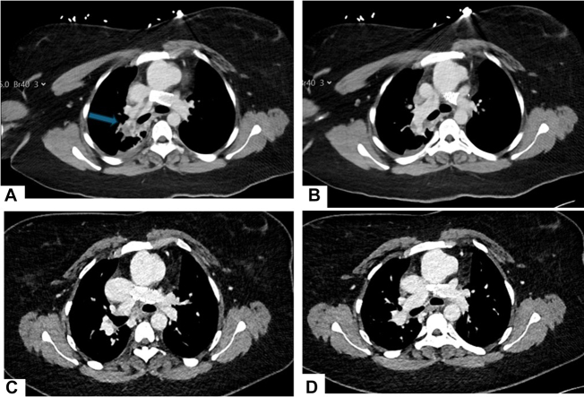
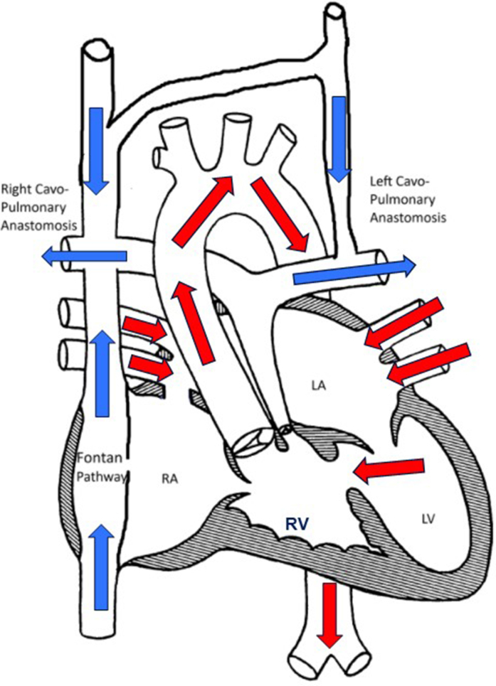
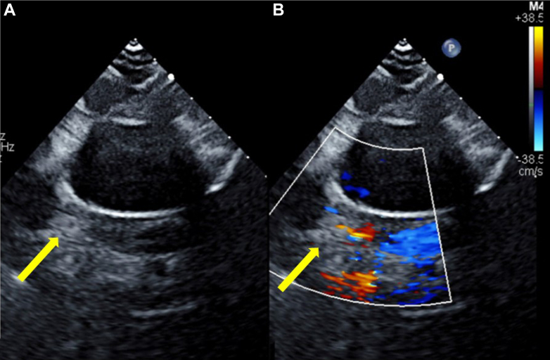
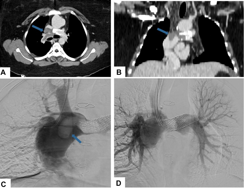
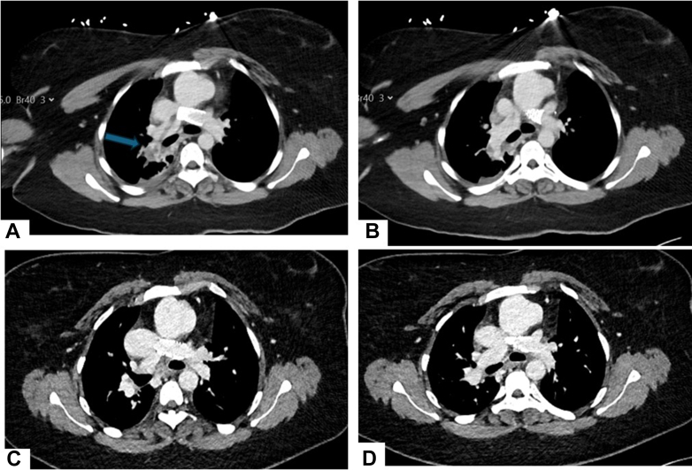
Case summary: A 26-year-old woman at 10 weeks gestation with a lateral tunnel Fontan experienced acute onset of dyspnea and dizziness. Cardiac CT demonstrated an occlusive pulmonary embolism within the Fontan circulation. The patient underwent pulmonary thrombectomy and catheter-directed thrombolysis without complications. A surveillance cardiac CT 3 months after her discharge demonstrated complete resolution of the thrombus, and the patient is at baseline functional capacity.
Discussion: Patients with Fontan circulation and pregnancy have a heightened risk for both thromboembolism and bleeding; anticoagulation strategies are thus not standardized and are tailored to each individual's case.
Take-home messages: To our knowledge, this is the first reported case of pulmonary embolism during the antepartum period in a patient with Fontan palliation who was subsequently treated with pulmonary thrombectomy and catheter-directed thrombolysis. Multimodality imaging evaluation was critical for rapid diagnosis and interventional planning.
Keywords: 3-dimensional imaging; Fontan; congenital heart disease; echocardiography; right-sided catheterization; thrombus.
Published by Elsevier Inc.
Conflict of interest statement
Funding Support and Author Disclosures The authors have reported that they have no relationships relevant to the contents of this paper to disclose.
Figures
References
-
- Rychik J., Atz A.M., Celermajer D.S., et al. Evaluation and management of the child and adult with Fontan circulation: a scientific statement from the American Heart Association. Circulation. 2019;140(6):e234–e284. - PubMed
-
- Heidendael J.F., Engele L.J., Bouma B.J., et al. Coagulation and anticoagulation in Fontan patients. Can J Cardiol. 2022;38(7):1024–1035. - PubMed
-
- Montanaro C., Boyle S., Wander G., et al. Pregnancy in patients with the Fontan operation. Eur J Prev Cardiol. 2024;31(11):1336–1344. - PubMed
-
- Cauldwell M., Steer P.J., Bonner S., et al. Retrospective UK multicentre study of the pregnancy outcomes of women with a Fontan repair. Heart. 2018;104(5):401–406. - PubMed
Publication types
LinkOut - more resources
Full Text Sources
