. 2025 Jun 20;45(6):1131-1142.
doi: 10.12122/j.issn.1673-4254.2025.06.03.

[ATF3 regulates inflammatory response in atherosclerotic plaques in mice through the NF-κB signaling pathway]

[Article in Chinese]
Affiliations

[ATF3 regulates inflammatory response in atherosclerotic plaques in mice through the NF-κB signaling pathway]

[Article in Chinese]
Bing Xia et al. Nan Fang Yi Ke Da Xue Xue Bao. .

Abstract

Objectives: To investigate the role of activating transcription factor 3 (ATF3) in atherosclerotic plaques for regulating inflammatory responses during atherosclerosis (AS) progression.

Methods: Human coronary artery specimens from autopsy cases were examined for ATF3 protein expression and localization using immunofluorescence staining and Western blotting. Apolipoprotein E-deficient (ApoE-/-) mouse models of AS induced by high-fat diet (HFD) feeding for 12 weeks were subjected to tail vein injection of adeno-associated virus serotype 9 (AAV9) to knock down ATF3 expression. After an additional 5 weeks of HFD feeding, the mice were euthanized for analyzing structural changes of the aortic plaques, and the expression levels of ATF3, inflammatory factors (CD45, CD68, IL-1β, and TNF-α), and NF-κB pathway proteins (P-IKKα/β and P-NF-κB p65) were detected. In the cell experiment, THP-1-derived foam cells were transfected with an ATF3-overexpressing plasmid or an ATF3-specific siRNA to validate the relationship between ATF3 and NF‑κB signaling.

Results: In human atherosclerotic plaques, ATF3 expression was significantly elevated and partially co-localized with CD68. ATF3 knockout in ApoE-/- mice significantly increased aortic plaque volume, upregulated the inflammatory factors, enhanced phosphorylation of the NF‑κB pathway proteins, and increased the expressions of VCAM1, MMP9, and MMP2 in the plaques. In THP-1-derived foam cells, ATF3 silencing caused activation of the NF‑κB pathway, while ATF3 overexpression suppressed the activity of the NF-κB pathway.

Conclusions: AS promotes ATF3 expression, and ATF3 deficiency exacerbates AS progression by enhancing plaque inflammation via activating the NF-κB pathway, suggesting the potential of ATF3 as a therapeutic target for AS.

目的: 探讨转录激活因子3(ATF3)在动脉粥样硬化斑块内的表达及其调控炎症反应参与动脉粥样硬化(AS)进程的机制。方法: 收集尸检案例中的人冠状动脉标本,免疫荧光和Western blotting检测ATF3蛋白表达及分布;高脂饮食12周后的载脂蛋白E基因敲除(ApoE-/-)小鼠尾静脉注射9型腺相关病毒(AAV9)敲低ATF3的表达,继续高脂喂养5周构建ATF3基因敲除的动脉粥样硬化ApoE-/-小鼠模型。麻醉处死后观察主动脉斑块结构改变,检测斑块内ATF3、炎症相关因子及NF-κB信号通路蛋白表达改变,并在体外构建ATF3的过表达质粒及siRNA干预THP-1诱导的泡沫细胞模型验证ATF3与NF-κB信号通路间关系。结果: 在人冠状动脉粥样硬化斑块内,ATF3的表达增高(P<0.05),且与CD68呈部分共表达;在ATF3基因敲除后,小鼠的主动脉斑块体积增大(P<0.05),斑块内炎症相关因子(CD45、CD68、IL-1β、TNF-α)表达增强(P<0.05),NF-κB信号通路相关蛋白(P-IKKα/β、P-NF-κB p65)表达增高(P<0.05),VCAM1、MMP9及MMP2表达增强(P<0.05);在离体THP-1细胞实验中验证了沉默ATF3后NF-κB信号通路进一步激活,而过表达ATF3后NF-κB信号受到抑制。结论: 动脉粥样硬化诱导ATF3的表达,ATF3的缺乏会促进AS的发生,增强斑块内炎症反应,其机制可能是通过进一步激活NF-κB信号通路导致,ATF3可能是AS潜在的治疗靶点。.

Keywords: activating transcription factor 3; atherosclerosis; inflammatory response; nuclear factor-κB; sudden cardiac death.


Figures
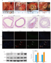
图1
图1
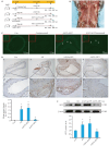
人冠状动脉实验 Fig.1 Examinations of human coronary arteries. A: Human coronary artery tissue specimens. B: Microscopic observation of coronary atherosclerosis (AS; HE staining, scale bar=1 mm). C: Partial co-localization of ATF3 (red) and CD68 (green) in the plaques (arrow,scale bar=50 μm). D: Western blotting for detecting ATF3 and CD68 expressions in the plaques. All experiments were repeated at least 3 times, and data are presented as Mean±SD (n=20). *P<0.05 vs Con group.
图2
图2
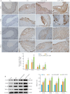
ApoE -/-小鼠ATF3基因敲除模型鉴定 Fig.2 Verification of ATF3 gene knockout in ApoE -/- mice. A: Schematic diagram of mouse grouping and treatment. B: Observation of a mouse aorta. The yellow lines indicate the sampling site, and the red box indicates the tissues used for preparing frozen sections or paraffin sections. C: Expression of eGFP in the aortic vessels. After masking autofluorescence in the mouse aortas with pontamine sky blue, no fluorescence signals were detected in the vessels in the control group, but eGFP expression (arrows) was observed in the aortic plaques in AAV9-eGFP group. D: Immunohistochemical staining for detecting distribution of ATF3 expression in the aorta (a1-d1 are enlarged images of the boxed areas in a-d. Scale bar=50 μm, n=6). E: Western blotting for detecting aortic ATF3 protein content (n=6). Data are presented as Mean±SD. *P<0.05 vs Con group; # P<0.05 vs AS or AAV9-eGFP group.
图3
图3
ATF3基因敲除后ApoE-/-小鼠主动脉斑块改变 Fig.3 Changes in mouse aortic plaques after ATF3 gene knockout. A: Gross oil red O staining of the aorta showing lipid deposition in the total vascular area (n=6). B: Oil red O staining of cross-sections within the plaque area showing lipid deposition in the aortic lumen (n=3). C: HE staining of the aortic plaque area (n=6). Scale bar=50 μm. Data are presented as Mean±SD. *P<0.05 vs Con group, # P<0.05 vs AS or AAV9-eGFP group.
图4
图4
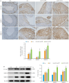
ATF3基因敲除后斑块内炎症因子表达增强 Fig.4 ATF3 gene knockout increases expressions of inflammatory factors in aortic plaques of the mice. A: Immunohistochemical staining for detecting aortic CD45, CD68, IL-1β and TNF-α protein expressions and MOD value statistics (scale bar=50 μm, n=6). B: Western blotting of aortic CD45, CD68, IL-1β and TNF-α protein expressions (n=6). Data are presented as Mean±SD. *P<0.05 vs Con group, # P<0.05 vs Con, AS or AAV9-eGFP group.
图5
图5
ATF3基因敲除后斑块内VCAM1、MMP-2和MMP-9蛋白增强 Fig. 5 ATF3 knockout increases expressions of VCAM1, MMP-2 and MMP-9 in aortic plaques of the mice. A: Immunohistochemical staining for detecting aortic VCAM1, MMP-2 and MMP-9 protein expressions and MOD value statistics (scale bar=50 μm, n=6). B: Western blotting for detecting aortic VCAM1, MMP-2 and MMP-9 protein expressions (n=6). Data are presented as Mean±SD. *P<0.05 vs Con group, # P<0.05 vs AS or AAV9-eGFP group.
图6
图6
ATF3基因敲除后NF-κB信号通路激活 Fig.6 ATF3 knockout activates the NF-κB signaling pathway in aortic plaques of the mice. A: Immunohistochemical staining for detecting aortic p-IKKα/β and p-NF-κB P65 protein expressions and MOD value statistics (scale bar=50 μm, n=6). B: Western blotting for detecting aortic IKKα/β, p-IKKα/β, NF-κB P65 and p-NF-κB P65 expressions (n=6). Data are presented as Mean±SD. *P<0.05 vs Con group, # P<0.05 vs AS or AAV9-eGFP group.
图7
图7
细胞实验结果 Fig.7 Cell experiment results. A: THP-1 cells, macrophages and foam cells stained with oil red O to identify lipid deposition (scale bar=50 μm). B: Western blotting for detecting ATF3 silencing efficiency and IKKα/β, p-IKKα/β, NF-κB P65, and p-NF-κB P65 protein expressions. C: Western blotting for detecting ATF3 overexpression efficiency and IKKα/β, p-IKKα/β, NF-κB P65, and p-NF-κB P65 protein expressions. Data are presented as Mean±SD. *P<0.05 vs PMA group, # P<0.05 vs PMA+Ox-LDL, PMA+Ox-LDL+Lip3000 or PMA+Ox-LDL+OE-vector group.

References

    1. Grandhi GR, Mszar R, Vahidy F, et al. Sociodemographic disparities in influenza vaccination among adults with atherosclerotic cardiovascular disease in the United States[J]. JAMA Cardiol, 2021, 6(1): 87-91. - PMC - PubMed
    1. Döring Y, van der Vorst EPC, Weber C. Targeting immune cell recruitment in atherosclerosis[J]. Nat Rev Cardiol, 2024, 21(11): 824-40. doi: 10.1038/s41569-024-01023-z - DOI - PubMed
    1. Akazawa H. Mechanisms of cardiovascular homeostasis and pathophysiology: from gene expression, signal transduction to cellular communication[J]. Circ J, 2015, 79(12): 2529-36. doi: 10.1253/circj.cj-15-0818 - DOI - PubMed
    1. Ghigo A, Laffargue M, Li MC, et al. PI3K and calcium signaling in cardiovascular disease[J]. Circ Res, 2017, 121(3): 282-92. doi: 10.1161/circresaha.117.310183 - DOI - PubMed
    1. Wang XW, Yan KY, Wen CF, et al. Simvastatin combined with resistance training improves outcomes in patients with chronic heart failure by modulating mitochondrial membrane potential and the Janus kinase/signal transducer and activator of transcription 3 signaling pathways[J]. Cardiovasc Ther, 2022, 2022: 8430733. doi: 10.1155/2022/8430733 - DOI - PMC - PubMed

Publication types

LinkOut - more resources