. 2025 Jun 28;13(1):134.
doi: 10.1186/s40478-025-02024-w.

Ultra-low-input cell-free DNA sequencing for tumor detection and characterization in a real-world pediatric brain tumor cohort

Affiliations

Ultra-low-input cell-free DNA sequencing for tumor detection and characterization in a real-world pediatric brain tumor cohort

Tom T Fischer et al. Acta Neuropathol Commun. .

Abstract

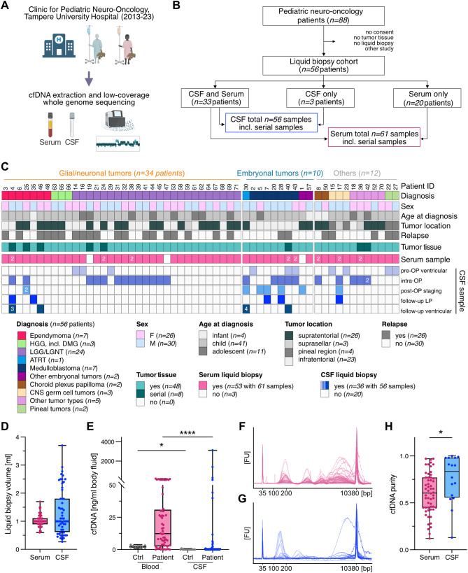
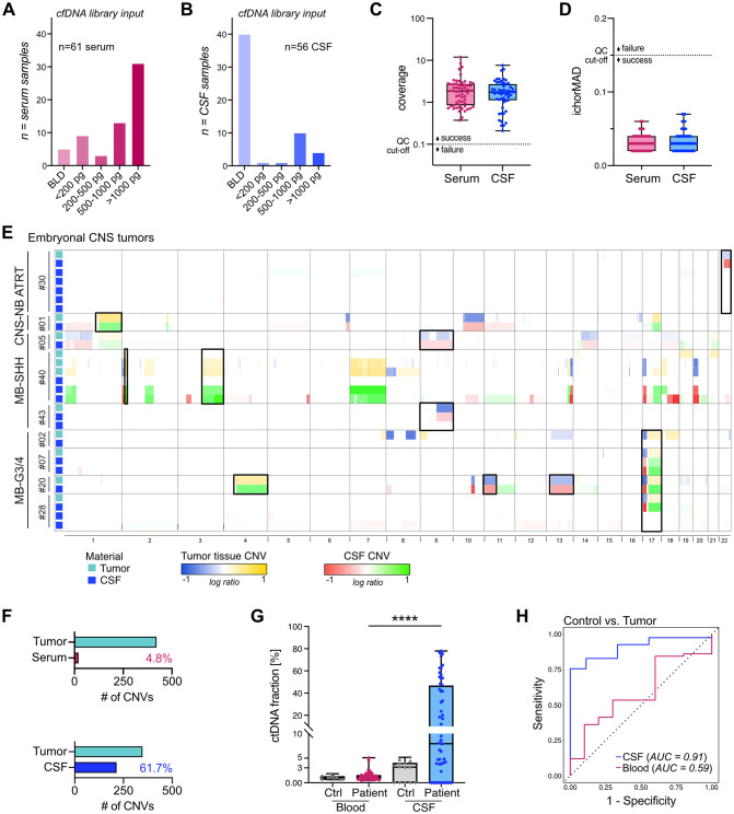
Molecular profiling of pediatric central nervous system (CNS) tumors has important clinical utility for guiding diagnostic and therapeutic strategies. Cell-free DNA (cfDNA) from liquid biopsies has been used for minimally invasive tumor profiling and longitudinal disease assessment in adult oncology and pediatric hematology. However, in pediatric neuro-oncology, low cfDNA yields pose a major barrier to translating these assays from bench to bedside. Here, we implemented a low-coverage whole genome sequencing (lcWGS) assay for picogram-level cfDNA inputs and applied it to liquid biopsies from a sizeable, population-based, cross-entity pediatric CNS tumor cohort (n = 56 patients). Applying this protocol, cfDNA whole genome profiles were successfully acquired from all liquid biopsy samples (n = 61/61 serum, n = 56/56 CSF, 100%). Based on copy number variations (CNVs), circulating-tumor DNA (ctDNA) was detected in 2/61 serum (3%) and in 25/56 CSF (45%) samples across various brain tumor entities. The integration of cfDNA results with clinical data demonstrated the utility of CSF lcWGS as a biomarker assay at diagnosis to distinguish cancerous from non-cancerous pineal region lesions (n = 6 patients). Additionally, serial CSF assessment in n = 9 patients (n = 29 CSF samples) enabled minimally invasive disease monitoring, with the added value of molecular profile availability in n = 4/6 (67%) patients at relapse. Proof-of-concept data show the feasibility of serial CSF lcWGS to reveal tumor evolution, tumor heterogeneity and potential therapeutic vulnerabilities in a case of medulloblastoma and germ cell tumor. Our study underscores the clinical utility of a robust lcWGS-based liquid biopsy assay optimized for low-input samples. We identify use-cases for implementing liquid biopsies in the clinical management of pediatric CNS tumor patients and provide a strong rationale for integration into future trials.

Keywords: Cell-free DNA; Cerebrospinal fluid liquid biopsies; Circulating-tumor DNA; Copy number variation profiling; Longitudinal molecular biomarkers; Low-coverage whole genome sequencing; Low-input samples; Minimally-invasive disease monitoring; Pediatric neuro-oncology; Tumor evolution.


Conflict of interest statement

Declarations. Ethics approval and consent to participate: The study was approved by the Finnish Committee of Ethics of Tampere University Hospital (ethics approval ID: R13050). Written, informed consent was obtained from all patients and/or their legal representatives. Consent for publication: Written informed consent for the publication was obtained from all study participants, their parents, or legal guardians. Competing interests: S.M.P.: scientific advisory board at BioSkryb. M.S., S.M.P., and F.S.: co-founders and shareholders of Heidelberg Epignostix GmbH. MS has become full-time employee of Heidelberg Epignostix GmbH since July 2024.

Figures

Fig. 1
Fig. 1
Overview of the pediatric neuro-oncology liquid biopsy cohort. (A) Schematic illustration of the study workflow. (B) Consort diagram delineating the composition of the study cohort and the liquid biopsy sample distribution. (C) Summary of the liquid biopsy study cohort (n = 56 cases), annotated with clinical information, biomaterials, and analyses performed. Numbers in cells denote sample size if > 1 liquid biopsy per sample type. (D) Available liquid biopsy volumes in ml. n = 61 serum (median = 1); n = 56 CSF (median = 1). (E) cfDNA concentration in ng/ml body fluid, n = 10 ctrl blood (median = 2.34); n = 61 patient blood ( median = 12.37); n = 14 ctrl CSF ( median = 0), n = 56 patient CSF ( median = 0). (F-G) Size distribution profiles of serum cfDNA and CSF cfDNA samples, respectively. (H) cfDNA purity quantified as cfDNA (1.-3. peak) over total DNA concentration (50–7000 bp) per sample, n = 56 serum, n = 16 CSF, samples with no measurable cfDNA excluded
Fig. 2
Fig. 2
lcWGS-based copy number profiling from minimal amounts of cell-free DNA. (A-D) Examples of lcWGS-derived CNV profiles for cfDNA input amounts on a range from nanograms to picograms demonstrating robust assay performance. (A) Patient #20 diagnosed with medulloblastoma group 3/4. CSF: collected intra-operatively, 1 ng of cfDNA used for library preparation. Serum: collected prior to surgery, 1 ng of cfDNA used for library preparation. (B) Patient #04 diagnosed with relapsed ependymoma PFA. CSF: collected via ventricular shunt during follow-up. 200 pg of cfDNA used for library preparation. (C) Patient #08 diagnosed with choroid plexus papilloma. CSF: collected intra-operatively, 100 pg of cfDNA used for library preparation. Serum: collected prior to surgery, 100 pg of cfDNA used for library preparation. (D) Patient #01 diagnosed with CNS neuroblastoma, FOXR2-activated. CSF: collected via lumbar puncture during post-operative staging. The cfDNA concentration was below the limit of detection, 10 µl of the 50 µl cfDNA isolate were used for library preparation
Fig. 3
Fig. 3
lcWGS-based tumor detection in pediatric CNS tumor liquid biopsies. (A-B) Bar graphs depicting cfDNA input amounts used for lcWGS library preparation for serum (A) and CSF (B) samples. BLD = below limit of detection. (C) Box plot showing sequencing coverage per sample, n = 61 serum (median = 1.83), n = 56 CSF (median = 1.74). All samples (117/117, 100%) passed the quality control threshold for a minimum coverage of > 0.1x. Y-axis was log10 transformed. (D) Box plot showing ichorMAD score per sample, n = 61 serum (median = 0.03), n = 56 CSF (median = 0.03). All samples (117/117, 100%) passed the quality control threshold for an ichorMAD score of < 0.15. MAD = median absolute deviation. (E) Genome-wide CNV profiles of CSF and matched tumor tissue samples for embryonal CNS tumors. Hallmark cytogenic events are highlighted with black boxes. CNS-NB = CNS neuroblastoma, FOXR2-activated. (F) Detection rate of tumor-derived CNVs in liquid biopsies across the study cohort: serum 20/419 (4.8%), CSF 214/347 (61.7%). In 9 of 56 patients, the tumor did not show CNVs. (G) Calculated ctDNA fraction in [%], n = 10 ctrl blood (median = 1%); n = 61 patient blood (median = 1%); n = 14 ctrl CSF (median = 3%), n = 56 patient CSF (median = 8%). (H) ROC curve for detecting tumor vs. control samples with a ctDNA fraction cut-off of 3% for serum and 5% for CSF. Only including samples where matched tumor has CNVs and liquid biopsy ctDNA fraction is > 0. n = 9 ctrl CSF, n = 41 patient CSF, n = 10 ctrl blood, n = 58 patient blood. ROC = receiver operating characteristic
Fig. 4
Fig. 4
Diagnostic utility of CSF liquid biopsies in pineal region tumors. (A) Oncoprint summarizing cohort of patients with pineal region involvement (n = 6 patients, n = 8 CSF samples). ctDNA positivity showed high biomarker accuracy in differentiating benign from malignant pineal region tumors. (B) Examples of MRI scans for two malignant pineal masses (case #15 germinoma, #22 pineoblastoma) and two benign pineal masses (case #26, #27). (C) CNV profiles derived from CSF and tissue biopsy for patient #27, diagnosed with a benign pineal region tumor. CSF collected pre-operatively from the third ventricle. (D) CNV profiles derived from CSF and resected tissue for patient #22, diagnosed with pineoblastoma. CSF collected pre-operatively from the third ventricle. (E) Genome-wide CNV profiles of CSF for all cases with pineal region involvement. (F) Schematic flowchart delineating potential implementation of cfDNA lcWGS into diagnostic work-up for pineal region masses. Abbreviations: AFP = alpha-1 fetoprotein, β-HCG = beta human chorionic gonadotropin, GCT = germ cell tumor, PB = pineoblastoma
Fig. 5
Fig. 5
Minimally invasive surveillance strategy via longitudinal CSF liquid biopsies. (A-B) Clinical timelines alongside CNV profiles, #X in timeline indicates number of days since diagnosis. (A) For patient #07, diagnosed with relapsed MB-G3/4, CSF during follow-up shows molecular relapse. (B) For patient #43, diagnosed with MB-SHH, CSF collected during suspected progression based on MRI was negative. (C) Serial trajectories of CSF ctDNA status, annotated by clinical time points, MRI results, and CSF cytology. Follow-up times for patients with no evidence of disease: #43: >2 years, #28: >1 year, #30: 9 months, #42: >1 year, #25: >1 year. (D) Schematic highlighting clinical utility of CSF-derived cfDNA as a biomarker, complementing MRI-based disease assessment. Abbreviations: EVD = external ventricular drain, GTR = gross total resection, LP = lumbar puncture, Rickham = ventricular reservoir
Fig. 6
Fig. 6
Assessment of tumor evolution using CSF-derived lcWGS profiles. (A) Clinical timeline alongside CNV profiles for patient #40 diagnosed with relapsed MB-SHH. (B) CNVs on Chr. 1 and 17 in matched tumor and CSF profiles displaying temporal evolution that was detected earlier in CSF than tissue (patient #40). (C) Clonal tree derived from Canopy analysis based on SNV and indel analysis from tumor, germline, and cfDNA CSF sequencing (patient #40). (D) Clonal composition for matched tumor and CSF samples displaying clonal evolution identified earlier in CSF than tissue (patient #40). (E) Clinical timeline combined with CNV profiles for patient #17 diagnosed with relapsed mixed GCT. (F) Evolution of broad and focal CNVs on indicated chromosomes and at indicated gene loci in tumor tissue and CSF over the course of the disease (patient #17). Abbreviations: Chr = chromosome, EVD = external ventricular drain, LP = lumbar puncture

References

    1. Siegel RL, Miller KD, Fuchs HE, Jemal A (2022) Cancer statistics, 2022. CA Cancer J Clin 72(1):7–33. 10.3322/caac.21708 - PubMed
    1. Ostrom QT, Price M, Ryan K, Edelson J, Neff C, Cioffi G et al (2022) CBTRUS statistical report: pediatric brain tumor foundation childhood and adolescent primary brain and other central nervous system tumors diagnosed in the united States in 2014–2018. Neuro Oncol 24(Suppl 3):iii1–iii38. 10.1093/neuonc/noac161 - PMC - PubMed
    1. Capper D, Jones DTW, Sill M, Hovestadt V, Schrimpf D, Sturm D et al (2018) DNA methylation-based classification of central nervous system tumours. Nature 555(7697):469–474. 10.1038/nature26000 - PMC - PubMed
    1. Gröbner SN, Worst BC, Weischenfeldt J, Buchhalter I, Kleinheinz K, Rudneva VA et al (2018) The landscape of genomic alterations across childhood cancers. Nature 555(7696):321–327. 10.1038/nature25480 - PubMed
    1. Louis DN, Perry A, Wesseling P, Brat DJ, Cree IA, Figarella-Branger D et al (2021) The 2021 WHO classification of tumors of the central nervous system: a summary. Neuro Oncol 23(8):1231–1251. 10.1093/neuonc/noab106 - PMC - PubMed

Publication types

MeSH terms

LinkOut - more resources