. 2025 Jun 28;74(8):254.
doi: 10.1007/s00262-025-04098-w.

Simultaneous TGF-β and GITR pathway modulation promotes anti-tumor immunity in glioma

Affiliations

Simultaneous TGF-β and GITR pathway modulation promotes anti-tumor immunity in glioma

Daniela Lorizio et al. Cancer Immunol Immunother. .

Abstract

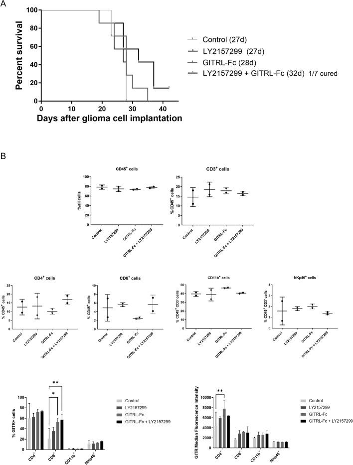
The immunosuppressive tumor microenvironment of glioblastoma limits the effectiveness of most immunotherapies. Transforming growth factor (TGF)-β signaling drives tumor progression and prevents effective T cell activity. Notably, both regulatory T cells (Tregs) and effector T cells within glioblastoma and other tumors express high levels of the immune checkpoint receptor, glucocorticoid-induced tumor necrosis factor receptor (GITR), which modulates T cell activation and function. Combining GITR agonism with TGF-β inhibition may therefore offer a compelling approach to restore anti-tumor immunity. We evaluated the combined effects of TGF-β inhibition and GITR modulation using two different GITR agonists in syngeneic mouse glioma models. GITR modulation enhanced T cell activation, as shown by increased cytokine secretion and effector T cell proliferation in vitro. Combining GITR modulation with TGF-β inhibition amplified these effects, resulting in significantly stronger immune cell-mediated tumor cell killing compared to single-agent treatments. Combination therapy improved survival of glioma-bearing mice, with a higher fraction of long-term survivors compared to monotherapy. Surviving mice resisted tumor re-challenge, indicating durable adaptive immunity. In summary, dual targeting of TGF-β and GITR pathways synergistically enhances anti-tumor immunity in glioblastoma. This novel combination strategy demonstrates clinical potential by addressing the limitations of existing immunotherapies and offering a promising approach for durable and effective glioblastoma treatment.

Keywords: GITRL; Glioblastoma; Immunosuppression; Immunotherapy; Microenvironment.


Conflict of interest statement

Declarations. Conflict of interest: MW has received research grants from Novartis, Quercis and Versameb, and honoraria for lectures or advisory board participation or consulting from Anheart, Bayer, Curevac, Medac, Neurosense, Novartis, Novocure, Orbus, Pfizer, Philogen, Roche, and Servier. PR has received honoraria for lectures or advisory board participation from Alexion, Bristol-Myers Squibb, Boehringer Ingelheim, CDR-Life, Debiopharm, Galapagos, Laminar, Midatech Pharma, Novartis, Novocure, OM Pharma, QED, Roche, Sanofi, and Servier and research support from Merck Sharp and Dohme and TME Pharma. DL, MS, JF, and AE have nothing to disclose. Ethics approval: All animal experiments were authorized by the Veterinary Office of the Canton of Zurich.

Figures

Fig. 1
Fig. 1
TGF-β stimulation and inhibition modulate immune checkpoint receptor expression on splenocytes. Splenocytes were exposed to hr-TGF-β2 (10 ng/ml), SD208 (1 µM), DMSO (control), or left untreated for 48 h, and immune checkpoint receptor expression was assessed by flow cytometry. Ratios of the median fluorescence intensity (MFI) of the specific antibody (blue) to its corresponding isotype control antibody (red) are shown in all panels
Fig. 2
Fig. 2
Simultaneous targeting of TGF-β and immune checkpoint receptors enhances T cell activation in syngeneic glioma co-cultures. A. SMA-560, SMA-540, GL-261, or CT2A glioma cells were co-cultured with syngeneic VM/Dk or C57BL/6-derived polyclonally activated splenocytes and exposed to a TGF-βRI inhibitor (SD208, 1 µM) (open symbols) or not (filled symbols), and antibodies targeting the immune checkpoint receptors GITR, CD137, OX40, LAG3, or PD-1 (10 µg/ml) or combinations thereof for 24 h (E:T = 10:1). Intracellular IFN-γ and IL-2 production by splenocytes was detected by flow cytometry, distinguishing CD4⁺ and CD8⁺ T cell subsets. For each condition, data are shown as individual values representing the four glioma cell lines. Results are expressed as mean relative frequency ± SD and were normalized to untreated control conditions (set to 1). B. SMA-560 cells were co-cultured with syngeneic splenocytes and exposed to an anti-GITR antibody or GITRL-Fc (10 µg/ml), either alone or combined with SD208 (1 µM) for 24 h. The percentage of CD4⁺ and CD8⁺ T cells expressing intracellular IFN-γ and IL-2 is shown for each treatment. Data are expressed as mean ± SD. C. Co-cultures as in (B) were incubated for 48 h. The Treg cell population was identified by flow cytometry as CD4+ CD25+ FoxP3+ cells. Data are expressed as mean ± SD. Statistical significance was assessed via one-way ANOVA with Dunnett’s post hoc test for multiple comparisons (* p < 0.05; ** p < 0.01; *** p < 0.001)
Fig. 3
Fig. 3
Combined TGF-β inhibition and GITR agonism enhance T cell-mediated glioma cell killing. Polyclonally activated splenocytes were exposed to an agonistic anti-GITR antibody or the GITRL-Fc fusion protein (10 µg/ml), a TGF-βRI inhibitor (SD208, 1 µM), or their combination for 48 h. Syngeneic mouse glioma cells, pre-stained with the fluorescent dye PKH-26, were co-cultured with these splenocytes for 24 h (E:T ratio = 5:1 for SMA-560 and CT2A cells, 10:1 for SMA-540, and GL-261 cells). Tumor cell lysis was assessed by flow cytometry, with the percentage of dead glioma cells shown. Data are indicated as mean ± SD. Statistical significance was determined via one-way ANOVA with Tukey’s post hoc test for multiple comparisons (* p < 0.05; ** p < 0.01; *** p < 0.001)
Fig. 4
Fig. 4
Combined inhibition of TGF-β and activation of GITR signaling increase NK cell-mediated glioma cell killing. Murine NK cells were exposed to the GITRL-Fc fusion protein (10 μg/ml), a TGF-βRI inhibitor (SD208, 1 μM), or the combination thereof for 24 h. Subsequently, PKH-26-labeled mouse glioma cells were co-cultured with the NK cells at the indicated effector:target ratios. Tumor cell lysis was assessed by flow cytometry after 20 h, as shown in the histograms. Data are presented as mean ± SD. Statistical significance was determined by one-way ANOVA with Tukey’s post hoc test for multiple comparisons (* p < 0.05; ** p < 0.01; *** p < 0.001)
Fig. 5
Fig. 5
Co-targeting of TGF-β and GITR signaling in the syngeneic SMA-560 mouse glioma model increases the number of long-term surviving animals. A-C. Twenty thousand SMA-560 cells were intracranially inoculated into the right striatum of syngeneic VM/Dk mice. From day 5 onward, mice were treated with a TGF-βRI inhibitor (LY2157299, 150 mg/kg, daily oral gavage), an agonistic GITR antibody (3 mg/kg, i.p. at days 5, 7, and 9), GITRL-Fc (30 μg/dose, i.p. from day 5 to day 9), or the respective combinations. A. Tumor size was assessed by H&E staining of tumors from three pre-randomized mice per group, harvested when the first mouse in any group displayed neurological symptoms. The histogram shows individual tumor volumes, with bars representing the mean volume for each treatment group. B. CD3 immunohistochemical staining was performed on one brain section per mouse. The histogram shows the mean number of CD3 + cells in three different regions of interest from the same section. The black bar represents the mean value for each group. C. Representative images of H&E, CD3, CD11b, and CD45 staining on tumor brain sections from one mouse per group. Scale bars: 100 µm for H&E, 20 µm for other stains. D. Twenty thousand SMA-560 cells were intracranially inoculated into the right striatum of syngeneic VM/Dk mice. From day 5 onward, mice were treated with a TGF-βRI inhibitor (LY2157299, 150 mg/kg, daily oral gavage), GITRL-Fc (30 μg/dose, i.p. from day 5 to day 9), or both. Survival data are shown, including the number of long-term surviving mice. E. On day 50 post-tumor inoculation, long-term surviving mice from experiments in (D) and Suppl. Figure 3B were re-challenged with a second tumor inoculation in the contralateral hemisphere. Three naïve mice injected with tumor cells served as controls. Kaplan–Meier survival curves are shown
Fig. 6
Fig. 6
Dual targeting of TGF-β and GITR signaling improves survival and modulates the phenotype of tumor-infiltrating immune cells in glioma-bearing mice. A, B. CT2A glioma cells were intracranially injected into the right striatum of C57B/6 mice. Five days after tumor cell inoculation, mice were treated with a TGF-βRI inhibitor (LY2157299, 150 mg/kg, daily oral gavage), GITRL-Fc (30 μg/dose, i.p. from day 5 to day 9), or the combination. A. Survival is shown by Kaplan–Meier curves. B. Tumor-infiltrating immune cells were isolated from randomized early-stage glioma-bearing mice and analyzed by flow cytometry. Immune cell subsets were defined with the indicated cell surface markers. Each dot represents one tumor sample, and the black bars indicate the average within the same treatment group (upper panel). The lower panels show the fraction of GITR+ cells among these immune cell subsets and the median fluorescence intensity of GITR

Similar articles

References

    1. Weller M, van den Bent M, Preusser M et al (2021) EANO guidelines on the diagnosis and treatment of diffuse gliomas of adulthood. Nat Rev Clin Oncol 18:170–186 - PMC - PubMed
    1. Ostrom QT, Price M, Neff C, Cioffi G, Waite KA, Kruchko C, Barnholtz-Sloan JS (2023) CBTRUS statistical report: primary brain and other central nervous system tumors diagnosed in the United States in 2016–2020. Neuro Oncol 25:iv1–iv99. 10.1093/neuonc/noad149 - PMC - PubMed
    1. Martinez-Lage M, Lynch TM, Bi Y et al (2019) Immune landscapes associated with different glioblastoma molecular subtypes. Acta Neuropathol Commun 7:1–12 - PMC - PubMed
    1. Nduom EK, Wei J, Yaghi NK et al (2015) PD-L1 expression and prognostic impact in glioblastoma. Neuro-oncol 18:195–205 - PMC - PubMed
    1. Mangani D, Weller M, Roth P (2017) The network of immunosuppressive pathways in glioblastoma. Biochem Pharmacol 130:1–9. 10.1016/j.bcp.2016.12.011 - PubMed

MeSH terms

Substances

LinkOut - more resources