. 2025 Sep:59:102461.
doi: 10.1016/j.tranon.2025.102461. Epub 2025 Jun 27.

Circulating mucosa-associated invariant T cells are decreased and have impaired function in patients with diffuse large B-cell lymphoma

Affiliations

Circulating mucosa-associated invariant T cells are decreased and have impaired function in patients with diffuse large B-cell lymphoma

Jingru Liu et al. Transl Oncol. 2025 Sep.

Abstract

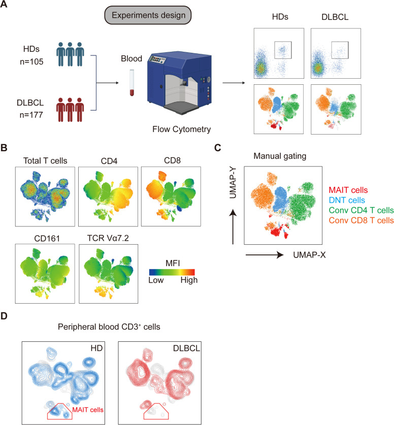
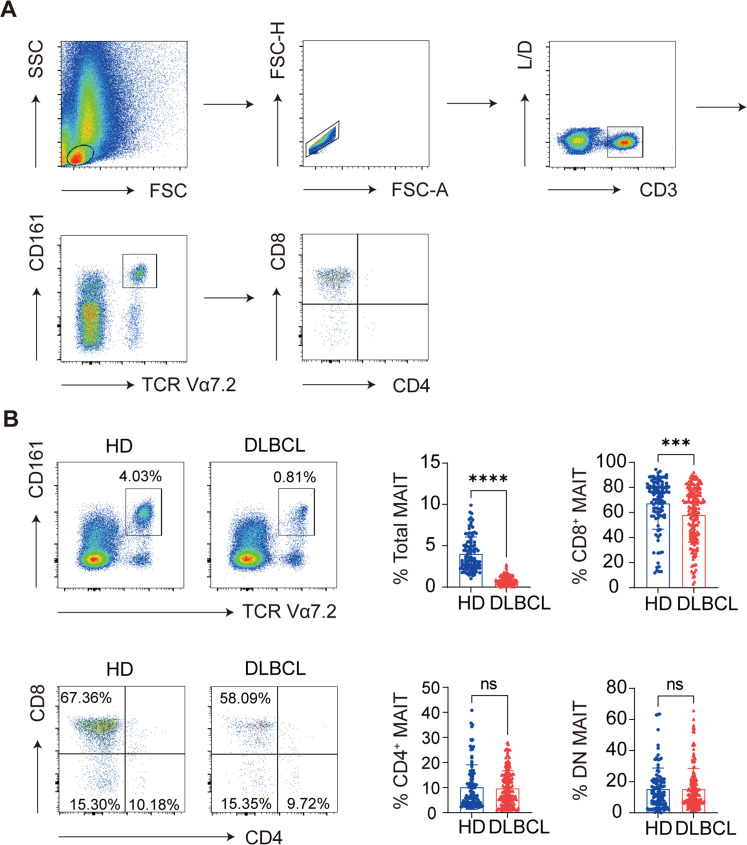
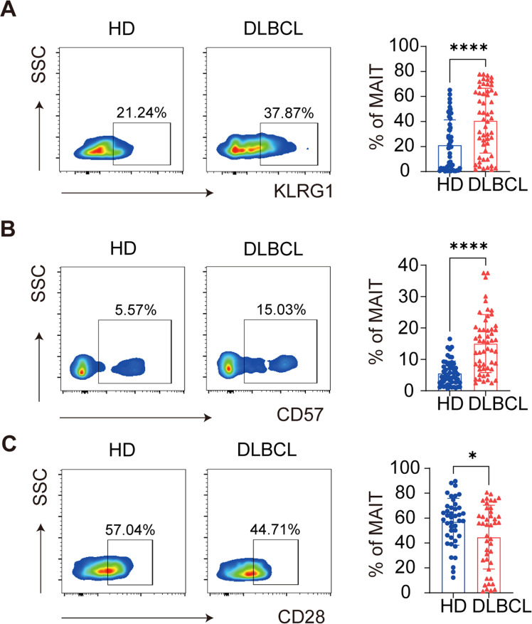
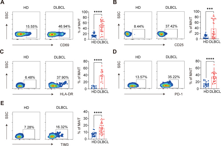
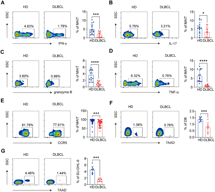
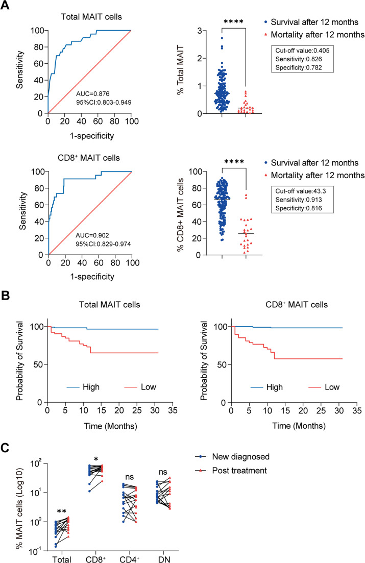
Mucosa-associated invariant T (MAIT) cells are associated with tumor immunity. However, their role in diffuse large B-cell lymphoma (DLBCL) remains unclear. Therefore, this study aimed to elucidate frequency and functional characteristics of circulating MAIT cells in DLBCL patients. The findings revealed a significant reduction in the frequency of circulating MAIT cells in DLBCL patients compared to age-matched healthy controls. Moreover, circulating MAIT cells from DLBCL patients exhibited proapoptotic and senescent phenotypes and demonstrated dysfunction, as evidenced by elevated expression of activation and exhaustion markers, including CD69, CD25, HLA-DR, PD-1, and Tim-3. MAIT cells derived from DLBCL produced lower levels of crucial anti-tumor cytokines, such as interferon-gamma, interleukin-17, tumor necrosis factor-α and granzyme B, suggesting impaired anti-tumor immunity. Additionally, MAIT cells from DLBCL patients showed diminished cytotoxicity against DLBCL cells compared to those from healthy donors. Notably, a lower frequency of circulating MAIT cells in patients with DLBCL was associated with poor prognosis. In summary, this study reveals reduced and impaired circulating MAIT cells in DLBCL patients, suggesting their importance in anti-lymphoma immunity.

Keywords: Diffuse large B-cell lymphoma; Dysfunction; Mucosa-associated invariant T cells; Prognosis.


Conflict of interest statement

Declaration of competing interest The authors declare that they have no known competing financial interests or personal relationships that could have appeared to influence the work reported in this paper.

Figures

Fig 1
Fig. 1
The frequency of circulating MAIT cells declines in patients with DLBCL at the new diagnosis. (A) Study design scheme. Flow cytometry was used to analyze mucosa-associated invariant T (MAIT) cells distribution in the peripheral blood of patients with large B-cell lymphoma (DLBCL) (n = 177) and healthy donors (HD) (n = 105). (B) Uniform Manifold Approximation and Projection (UMAP) plots of total live CD3+T cells in the peripheral blood showing the expression of the indicated markers. (C) UMAP plots of total live CD3+T cells overlaid with immune subsets identified by manual gating. (D) Colored according to the DLBCL (n = 23) and HD (n = 21) groups. CD3+ cells (n = 3000 per sample) were downsampled, barcoded according to the two groups, and concatenated. The red circle indicates the MAIT cell compartment.
Fig 2
Fig. 2
Distribution of different circulating MAIT cell subsets from patients with DLBCL and HDs (A) Flow cytometric gating strategy for mucosa-associated invariant T (MAIT) cells and their subpopulations. (B) Distribution and proportion of MAIT cells in the peripheral blood of patients with large B-cell lymphoma (DLBCL) (n = 177) and healthy donors (HD) (n = 105). (***P < 0.001, ****P < 0.0001; ns: no significance).
Fig 3
Fig. 3
Circulating MAIT cells in patients with DLBCL exhibit increased apoptosis and reduced proliferation. (A) Flow cytometric analysis and quantification of apoptotic circulating mucosa-associated invariant T (MAIT) cells from patients with large B-cell lymphoma (DLBCL) (n = 36) and healthy donors (HD) (n = 18). (B) Flow cytometric analysis and quantification of the Ki67 expression levels in circulating MAIT cells from patients with DLBCL (n = 12) and HD (n = 14). (C) Flow cytometric analysis and quantification of apoptotic circulating MAIT cells co-cultured with peripheral blood mononuclear cells (PBMC) (n = 17), DB cells (n = 23) and SU-DHL-6 cells (n = 13). ( ****P < 0.0001; ns: no significance).
Fig 4
Fig. 4
Circulating MAIT cells in patients with DLBCL are prone to senescence. (A) Flow cytometric analysis and quantification of KLRG1 expression levels in circulating mucosa-associated invariant T (MAIT) cells from patients with large B-cell lymphoma (DLBCL) (n = 50) and healthy donors (HD) (n = 52). (B) Flow cytometric analysis and quantification of CD57 expression levels in circulating MAIT cells from patients with DLBCL (n = 53) and HD (n = 52). (C) Flow cytometric analysis and quantification of CD28 expression levels in circulating MAIT cells from patients with DLBCL (n = 47) and HD (n = 44). (*P < 0.05, **** P < 0.0001).
Fig 5
Fig. 5
Circulating MAIT cells in patients with DLBCL exhibit an activated and exhausted phenotype. (A and B) Flow cytometry analysis and quantification of CD69 (A) and CD25 (B) expression levels in circulating mucosa-associated invariant T (MAIT) cells from patients with large B-cell lymphoma (DLBCL) (n = 30) and healthy donors (HD) (n = 20). (C) Flow cytometric analysis and quantification of HLA-DR expression in circulating MAIT cells from patients with DLBCL (n = 16) and HDs (n = 14). (D and E) Flow cytometric analysis and quantification of the expression levels PD-1 (D) and Tim-3 (E) in circulating MAIT cells from patients with DLBCL (n = 30) and HD (n = 20). (***P < 0.001, ****P < 0.0001).
Fig 6
Fig. 6
Circulating MAIT cells in patients with DLBCL exhibit lower cytokine levels and less cytotoxic ability. (A-E) Flow cytometric analysis and quantification of interferon-gamma (IFN-γ) (A), interleukin-17 (IL-17) (B), granzyme B (C), and tumor necrosis factor-alpha (TNF-α) (D) secretion from circulating mucosa-associated invariant T (MAIT) cells from patients with large B-cell lymphoma (DLBCL) (n = 15) and healthy donors (HD) (n = 15). (E) Flow cytometric analysis and quantification of the CCR5 expression levels in circulating MAIT cells from patients with DLBCL (n = 40) and HDs (n = 26). (F-G) Flow cytometric analysis and quantification of the cytotoxicity of MAIT cells from DLBCL. (DB cells, n = 8; SU-DHL-6 cells, n = 8) (* P < 0.05; *** P < 0.001, ****P < 0.0001;).
Fig 7
Fig. 7
Circulating MAIT cells are associated with improved overall survival. (A) Receiver operating characteristic curve (ROC) and cut-off values for the percentages of circulating total mucosa-associated invariant T (MAIT) cells and CD8+ MAIT cells to predict 12 months survival. (B) High percentages of circulating total MAIT and CD8+ MAIT cells predict improved overall survival (OS) in patients with large B-cell lymphoma (DLBCL). Patients with DLBCL with total MAIT cell frequency ≤ 40.5 % (n = 53), > 40.5 % (n = 117); patients with DLBCL with CD8+ MAIT cells frequency ≤ 43.3 % (n = 48), > 43.4 % (n = 122); (C) The alterations of circulating total MAIT cells and MAIT cell subsets in new diagnosed (n = 17) patients with DLBCL and post treatment (n = 17). (* P < 0.05, **P < 0.005, **** P < 0.0001; ns: no significance).

Similar articles

References

    1. Petley E.V., Koay H.F., Henderson M.A., et al. MAIT cells regulate NK cell-mediated tumor immunity. Nat. Commun. 2021;12(1):4746. - PMC - PubMed
    1. Liu Y., Zhang Q., Xing B., et al. Immune phenotypic linkage between colorectal cancer and liver metastasis. Cancer Cell. 2022;40(4):424–437. e5. - PubMed
    1. Ruf B., Bruhns M., Babaei S., et al. Tumor-associated macrophages trigger MAIT cell dysfunction at the HCC invasive margin. Cell. 2023;186(17):3686–3705. e32. - PMC - PubMed
    1. Garner L.C., Amini A., FitzPatrick M.E.B., et al. Single-cell analysis of human MAIT cell transcriptional, functional and clonal diversity. Nat. Immunol. 2023;24(9):1565–1578. - PMC - PubMed
    1. Pellicci D.G., Koay H.F., Berzins S.P. Thymic development of unconventional T cells: how NKT cells, MAIT cells and γδ T cells emerge. Nat. Rev. Immunol. 2020;20(12):756–770. - PubMed