. 2025 Jun 28;9(1):205.
doi: 10.1038/s41698-025-00997-4.

Explainable, federated deep learning model predicts disease progression risk of cutaneous squamous cell carcinoma

Affiliations

Explainable, federated deep learning model predicts disease progression risk of cutaneous squamous cell carcinoma

Juan I Pisula et al. NPJ Precis Oncol. .

Abstract

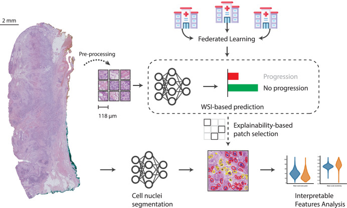
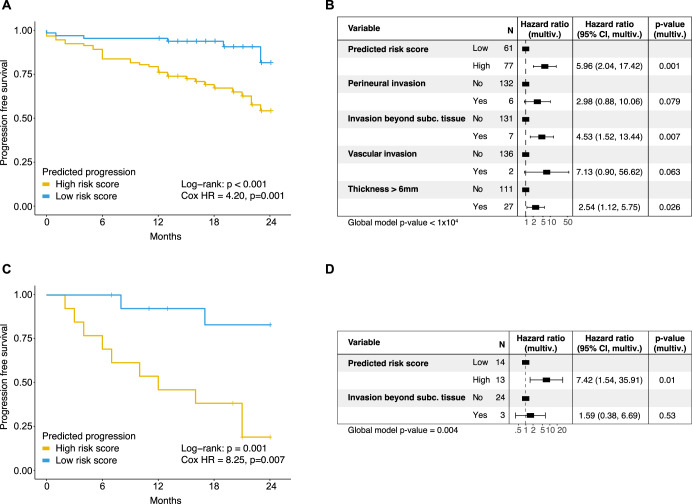
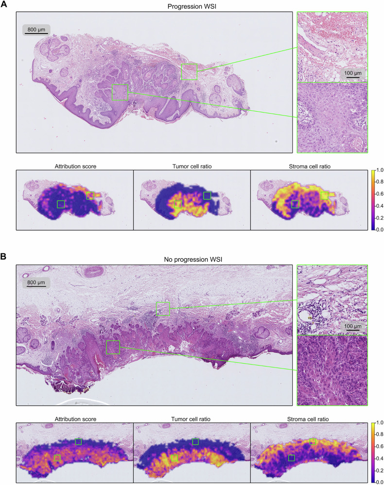
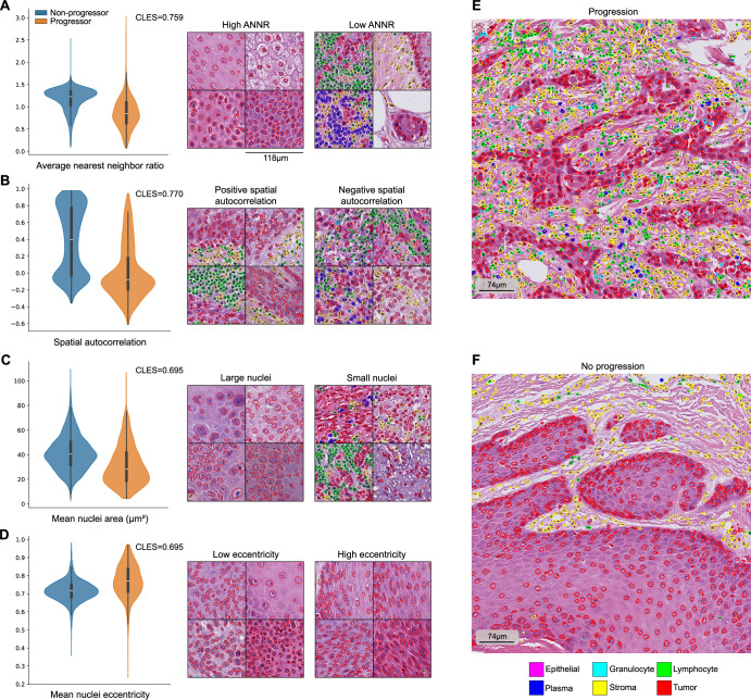
Predicting cancer patient disease progression is a key step towards personalized medicine and secondary prevention. Risk stratification systems based on clinico-pathological criteria aim to identify high-risk patients, but accurate predictions remain challenging. Deep learning models present new opportunities for patient risk prediction, yet their interpretability has been largely unexplored. We developed a transformer-based approach for predicting progression of cutaneous squamous cell carcinoma (cSCC) patients based on diagnostic histopathology tumor slides. Our initial model showed AUROC = 0.92 on a held-out test set, with average AUROC of 0.65 on external validation cohorts. To further increase generalizability and reduce potential privacy concerns, we trained the model in a federated manner across three clinical centers, reaching AUROC = 0.82 across all cohorts, with image-based risk scores achieving hazard ratios up to 7.42 (p < 0.01) in multivariable analyses. Through interpretability analysis, we identified spatial and morphological features predictive of progression, suggesting that tumor boundary information and tissue heterogeneity characterize progressive cSCCs. Trained exclusively on routine diagnostic slides and offering biological insights, our model can improve secondary prevention and understanding of cSCC while enabling deployment across clinical centers without administrative overheads or privacy concerns.


Conflict of interest statement

Competing interests: The Authors declare no Competing Non-Financial Interests but the following Competing Financial Interests: D.N. received financial support (speaker’s honoraria, advisory boards, travel expense reimbursements or grants) from Abbvie, Almirall, AstraZeneca, Biogen, Boehringer Ingelheim, Bristol-Myers-Squib, GlaxoSmithKline, Incyte, Janssen-Cilag, Kyowa Kirin, LEO Pharma, Lilly, L’Oreal/Cerave, MSD, Novartis, Pfizer, Regeneron and UCB Pharma. J.B. received research funding from Bayer and travel expenses from Merck KG and Bicycle Therapeutics outside the presented work. K.D. received financial support (speaker’s honoraria, advisory boards, travel expense reimbursements or grants) from Abbvie, Bristol-Myers-Squib, Novartis, and Pierre-Fabre.

Figures

Fig. 1
Fig. 1. We propose a WSI-based cutaneous Squamous Cell Carcinoma (cSCC) progression prediction model, trained on data from three medical centers using Federated Learning.
Beyond prediction, we investigate underlying biological features that influence our classifier. We do so by computing cellular-level features with aid of a nuclei segmentation model. We analyze these features in image regions detected as relevant for prediction outcome by Integrated Gradients, an input attribution algorithm for explainable deep neural networks.
Fig. 2
Fig. 2. ROC curves of the classifiers.
A WSI-based classifier trained exclusively on the Cologne cohort and tested on Munich and Bonn cohorts (AUROC = Area under the receiver operator curve). B Multivariate logistic regression model based on clinico-pathological parameters associated with progression risk in univariate analysis. Model trained and evaluated on the Cologne cohort. C Federated WSI-based classifier.
Fig. 3
Fig. 3. Comparison of federated and original deep learning models for survival prediction.
A, B Federated model trained on Bonn and Munich cases, applied to Cologne patients. C, D Original model trained on Cologne, applied to Bonn patients. A Progression-free survival of Cologne patients classified as high vs. low progression risk based on federated deep learning prediction (threshold: Youden index, HR from univariate Cox regression). B Multivariable Cox regression for n = 138 Cologne patients, integrating federated deep learning risk categories with clinical parameters. C Progression-free survival of Bonn patients classified using the original Cologne model (threshold: Youden index, HR from univariate Cox regression). D Multivariable Cox regression for n = 27 Bonn patients, combining deep learning risk categories with clinical parameters.
Fig. 4
Fig. 4. Slides and heatmaps of the patches’ classifier attribution score, tumor cell ratio, and stroma cell ratio.
A Slide of a progression patient, showing that the WSI-based classifier assigns higher importance to the region outside the tumor area (indicated by the tumor cell ratio heatmap). B Slide of a non-progression patient, where the high attribution area coincides with the tumor-cell populated areas. Colorbar indicates the slide-normalized heatmap values.
Fig. 5
Fig. 5. Four of the features of the tumor cells used in the analysis.
A−D show violin plots and segmented image patches that illustrate these values. In general, progression-associated tumor cells cluster together (A), interface with other cell types (B), and have smaller (C), eccentric nuclei (D). These effects are not just local to image patches, but they occur in larger regions, as shown in (E, F). The displayed CLES (Common Language Effect Size) values are indicated for the group with the largest mean. All features are significantly different in both groups, with p-values < 0.0001 using Mann–Whitney U-test.

References

    1. Winge, M. C. G. et al. Advances in cutaneous squamous cell carcinoma. Nat. Rev. Cancer23, 430–449 (2023). - PubMed
    1. Keim, U. et al. Incidence, mortality and trends of cutaneous squamous cell carcinoma in Germany, the Netherlands, and Scotland. Eur. J. Cancer Oxf. Engl. 1990183, 60–68 (2023). - PubMed
    1. Brantsch, K. D. et al. Analysis of risk factors determining prognosis of cutaneous squamous-cell carcinoma: a prospective study. Lancet Oncol.9, 713–720 (2008). - PubMed
    1. Schmults, C. D., Karia, P. S., Carter, J. B., Han, J. & Qureshi, A. A. Factors predictive of recurrence and death from cutaneous squamous cell carcinoma: a 10-year, single-institution cohort study. JAMA Dermatol.149, 541–547 (2013). - PubMed
    1. Thompson, A. K., Kelley, B. F., Prokop, L. J., Murad, M. H. & Baum, C. L. Risk factors for cutaneous squamous cell carcinoma recurrence, metastasis, and disease-specific death: a systematic review and meta-analysis. JAMA Dermatol152, 419–428 (2016). - PMC - PubMed

LinkOut - more resources