. 2025 Aug 4;198(4):kiaf269.
doi: 10.1093/plphys/kiaf269.

Insights into plastocyanin-cytochrome b6f complex formation: The role of plastocyanin phosphorylation

Affiliations

Insights into plastocyanin-cytochrome b6f complex formation: The role of plastocyanin phosphorylation

Yuval Milrad et al. Plant Physiol. .

Abstract

Plastocyanin (PC) is a copper-containing protein that acts as a mobile electron carrier in plants during photosynthesis. In this work, we investigated the role of PC phosphorylation in photosynthetic electron transfer, focusing on interactions with both cytochrome b6f (Cytb6f) and photosystem I (PSI) in Chlamydomonas reinhardtii. While the binding and electron transfer between PC and PSI are well characterized, the interaction between PC and Cytf remains less clear. Using chemical cross-linking combined with mass-spectrometry, we identified 2 potential binding models for PC and Cytf: "side-on" and "head-on." To evaluate electron transfer, we developed an in vitro system that allowed oxidized PC, formed via light-driven electron transfer at PSI, to reoxidize Cytf. Our data show that a phosphomimetic variant of PC, where phosphorylated PC S49 residue interacts with PetA-K188, displays faster Cytf oxidation, likely optimizing binding and electron transfer between PC and Cytf. Additionally, PC phosphomimetic variants exhibited slower transfer rates than the wild type, suggesting that phosphorylation also modulates PC's interaction with PSI. This regulation likely optimizes Cytf oxidation and electron transfer under conditions of low PC availability, such as during high light stress. Overall, PC phosphorylation appears to play a role in fine-tuning electron transfer between PSI, Cytb6f, and PC, thereby ensuring efficient photosynthesis in dynamic environmental conditions.


Conflict of interest statement

Conflict of interest statement. None declared.

Figures

Figure 1.
Figure 1.
A) Alignment-based comparison of relevant loops was conducted for PsaF and PC, using all known sequences (Uniprot.org) of plant (Streptophyta) and green algae (Chlorophyta) orders. Relevant residues are shown in bold. B, C, and D) Superposed structure of PC and PSI. Relevant acidic residues (facing the PsaF loop) are presented (blue sticks) as well as 2 serine residues (S10 and S49, yellow), which were shown to be phosphor-regulated. B, C) The formed salt bridges between PC and PSI, comparing plants (B, PDB: [6ZOO]) and algae (C, PDB: [7ZQE], [7ZQC]) systems. Nonconserved residues of PsaF (green) are highlighted (orange). D) Side view of PC from C. reinhardtii. Note that the copper cofactor is situated directly beneath the double tyrosine gateway, leading to the P700 reaction center (orange sticks). E) Structural illustration of Cytf (backbone yellow) from C. reinhardtii (PDB: [1Q90]), emphasizing its interaction plane and relevant residues (e.g. lysines, orange). F) Alignment-based comparison of relevant loops was conducted for petA (Cytf), using all known sequences of plant (Streptophyta) and green algae (Chlorophyta) orders. Relevant residues and regions are shown in bold. The illustration was generated using BioRender.com.
Figure 2.
Figure 2.
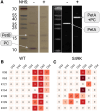
Recombinant PC was activated with EDC and NHS and cross-linked with purified His-tag Cytb₆f. SDS-PAGE fractionation of these samples showed cross-linked (NHS+) and noncross-linked (NHS−) bands in both Coomassie blue stain and PetA antibodies based on Western blots (size markers in white boxes were overlaid using the system's software; for full images, see Supplementary Fig. S1) A). Cross-linked samples of 3 biological replicas from WT strains were digested by trypsin and analyzed via MS; positive detections were summed. The results revealed an abundant PC:D59/D66 residues B). Although previous models predicted tight interaction between the southern loop of PC (D43, E44, D45, and D54) and PetA (K188 and K189), hardly any crosses were detected (WT). We therefore tested recombinant PC mutants with a point mutation at S49K (4 biological replicas), generating additional cutting site, and thus enabled a better detection of the relevant peptides C). The illustration was generated using BioRender.com.
Figure 3.
Figure 3.
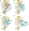
Presented are the models of complex formation between Cytf (yellow-orange) and algal PC (cyan). By superposing algal PC (PDB 7ZQC) to the NMR-based plant interaction model (PDB 2PCF) (presented in A, plant PC in gray50), we tested the distances between the negatively charged PC resides (blue) and positively charged Cytf residues (orange). In accordance with this model, we measured the distance between the metal cores of the proteins (PC-Cu/PetA-heme-Fe) and determined them to be 11.9 Å B). The second model C) takes into consideration the fact that algal PetA has an additional Lys at Position 121 and that it was cross-linked to PC:D59/D60. Additional interactions are observed between PC:D54 and PetA-K58/K65 (as was shown in the mutated S49K PC), PC:D43/E44, and PetA-K188/K65. In addition, PC:S49 seems to be in proximity of PetA-K188 and given a phosphorylation form might increase the stability of the complex formation. Core Cu–Fe distances are predicted to be 10.9 Å. The third model D) takes into consideration a possible interaction between PC:D60 and PetA-K164. This residue cannot be found in plants or Cyanobacteriota, but only in green algae. Since we observed such cross-linked peptides, we aligned the residues and postulated that the chances for this orientation to dominate the interaction mode increase under conditions in which PC:S10 is phosphorylated and PC:S49 remains unphosphorylated. Core Cu–Fe distances are predicted to be 10.9 Å, which should increase the electron transfer rate. In addition, other negative residues of PC are not in interaction, which should decrease the complexes strength. All models are available on Supplementary File S4. The illustration was generated using BioRender.com.
Figure 4.
Figure 4.
Purified PSI complexes were tested in JTS. A) Following a laser flash, maximal oxidation of P700+ centers were determined, followed by a double exponential re-reduction phase. The complexes were mixed with increasing PC concentration, and re-reduction rate was increased accordingly. The kinetics featured 2 phases of reduction, in which the initial reduction, below measurement resolution, was detected. The results show averaged plots of 3 biological replicas. To test the effects of phosphorylated PC variants on the kinetics of P700+ re-reduction, the experiment was conducted in the presence of recombinant PC variants B), in which S10 or S49 were replaced by either Ser (WT), Ala (S10A), Glu (S10E), or Asp (S49D). K2 values were calculated, using OriginLab (expDec2), for each concentration of PC, and results show averaged values of 3 biological replicas with SEM error bars. C) Box plots (center line, median; box limits, upper and lower quartiles; whiskers, 5 to 95 percentile; points, outliers) show the averaged K2 values of 1 to 6 µm PC (3 biological repetitions). Here, S10A showed no significant change, while both phosphor-mimicking mutations showed a slight decrease. Statistical analysis was conducted using a 1-way ANOVA with Dunnett multiple comparisons test. The illustration and statistical analysis were generated using BioRender.com.
Figure 5.
Figure 5.
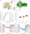
A) Schematic overview of the cyclic electron chain between cytochrome b6f (Cytb₆f, yellow) and PSI (green) via PC (cyan) and FDX (orange), using ascorbate (Asc, brown) as a reducing agent, as was described in our experimental setup. Kinetic measurements were conducted using JTS on isolated Cytb6f (2 µm) and PSI (300 nm) complexes, in the presence of 1 mm ascorbate and 2.5 mm MgCl2 at pH 7.0. B) Post laser flash P700 measurements were conducted, with increasing concentrations of FDX, in the presence of 1 µm PC (WT, solid red lines on a gradient scale). Before and after FDX additions, PC was added to illustrate the range of expected changes (dashed lines), with PC additions from 0.3 to 1.0 µm (gray gradient) and from 1.0 to 3.0 µm (blue gradient). C)  K2 values of P700⁺ re-reduction rates were calculated using OriginLab (expDec2) and presented as box plots (center line, median; box limits, upper and lower quartiles; whiskers, 5 to 95 percentile; points, outliers), in the presence/absence of FDX (FDX+/−), where FDX− was supplemented with methyl viologen (blue). The effect of Cytb₆f complexes was also tested (Cyt+, orange). Results show no significant differences between these treatments. Cytf oxidation kinetics were also tested, as complexes were exposed to 2 s of illumination (white background). D) Increasing FDX concentrations showed decreased oxidation kinetics (colored in red gradient), possibly due to a faster re-reduction of cytochrome complexes. E) Increasing PC concentrations (in the presence of 5 µm FDX, colored in blue gradient) featured an increase of the second light-phase net oxidation rate and a rapid dark re-reduction (gray background). The illustration was generated using BioRender.com.
Figure 6.
Figure 6.
A) P700+ re-reduction assay (3 biological replica, analysis was conducted as described in Fig. 5C) was conducted in the absence (blue) or presence (orange) of Cytb6f, using different PC mutants (WT, S10A, S10E, and S49D). Both S10E and S49D PC peptides showed an additional decrease in K2 values (2-way ANOVA with Bonferroni multiple comparisons test, considering 3 biological replicas of all tested samples; ** indicates P-values of smaller than 0.01), indicating a decrease of its apparent concentration and suggesting a stronger interaction with Cytf. B) Comparative Cytf reduction assay (as described in Fig. 5E), where samples were exposed to 2 s of illumination (white background), before the light was turned off. The graph shows relative averaged plots (based on oxidation at 2 s following light onset, colored as WT black, S10A blue, S10E red, and S49D yellow in B to D) of 3 biological replicas with SEM bars. C) Box plots (center line, median; box limits, upper and lower quartiles; whiskers, 5 to 95 percentile; points, outliers) of Cytf oxidation kinetics showed a significant faster oxidation when S49D PC was added (1-way ANOVA with Dunnett multiple comparisons test with; ** indicates P-values of smaller than 0.001). D) Dark re-reduction of Cytf showed an increased rate when PC:S49D was added, but not due to FDX additions. The graph shows averaged half-time values (OriginLab, expDec2) of 3 biological replicas with SEM bars. Statistical analysis was conducted using a 1-way ANOVA with Dunnett multiple comparisons test (for 3 biological repetitions). The illustration and statistical analysis were generated using BioRender.com.

Similar articles

References

    1. Allen JF. Protein phosphorylation in regulation of photosynthesis. Biochim Biophys Acta. 1992:1098(3):275–335. 10.1016/S0005-2728(09)91014-3 - DOI - PubMed
    1. Balparda M, Bouzid M, Martinez MDP, Zheng K, Schwarzländer M, Maurino VG. Regulation of plant carbon assimilation metabolism by post-translational modifications. Plant J. 2023:114(5):1059–1079. 10.1111/tpj.16240 - DOI - PubMed
    1. Bellafiore S, Barneche F, Peltier G, Rochaix J-D. State transitions and light adaptation require chloroplast thylakoid protein kinase STN7. Nature. 2005:433(7028):892–895. 10.1038/nature03286 - DOI - PubMed
    1. Bergner SV, Scholz M, Trompelt K, Barth J, Gäbelein P, Steinbeck J, Xue H, Clowez S, Fucile G, Goldschmidt-Clermont M, et al. STATE TRANSITION7-dependent phosphorylation is modulated by changing environmental conditions, and its absence triggers remodeling of photosynthetic protein complexes. Plant Physiol. 2015:168(2):618–634. 10.1104/pp.15.00072 - DOI - PMC - PubMed
    1. Buchert F, Hamon M, Gäbelein P, Scholz M, Hippler M, Wollman FA. The labile interactions of cyclic electron flow effector proteins. J Biol Chem. 2018:293(45):17559–17573. 10.1074/jbc.RA118.004475 - DOI - PMC - PubMed

MeSH terms

LinkOut - more resources