. 2025 Sep;24(9):e70154.
doi: 10.1111/acel.70154. Epub 2025 Jun 29.

Impaired Cytokine Secretion Contributes to Age-Dependent Immune Dysfunction in SARS Coronavirus Response and Is Restored by Young CD11b-Positive Cell Transfer

Affiliations

Impaired Cytokine Secretion Contributes to Age-Dependent Immune Dysfunction in SARS Coronavirus Response and Is Restored by Young CD11b-Positive Cell Transfer

Yu-Xuan Wu et al. Aging Cell. 2025 Sep.

Abstract

COVID-19 mortality disproportionately affects the elderly, yet the cellular and molecular factors contributing to age-related immune system remodeling remain unclear. Using SARS-CoV-derived ssRNA sequences, we modeled age-dependent immune responses in mice. Aged mice exhibited higher mortality and severe lung inflammation upon viral ssRNA challenge, mirroring clinical observations. We uncovered a pre-existing inflammatory state in aged mice, characterized by elevated baseline levels of specific immune cells and cytokines correlating with poor outcomes. Age-related immune dysfunction stemmed from impaired IRF7 signaling and defective SNARE-mediated cytokine secretion in CD11b+ cells. Notably, the adoptive transfer of young CD11b+ cells to aged mice exposed to SARS-CoV2 ssRNA reduced mortality, alleviated lung inflammation, and normalized cytokine profiles. These findings provide insights into age-related immune dysregulation during viral challenges and suggest potential therapeutic strategies for severe COVID-19 in the elderly.

Keywords: IRF7; SARS‐CoV; SNARE; aging; innate immunity.


Conflict of interest statement

The authors declare no conflicts of interest.

Figures
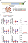
FIGURE 1
FIGURE 1
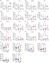
Comparison of SARS‐CoV‐derived GU‐rich ssRNA sequences and their hematological effects versus live SARS‐CoV‐2 BA.5 variant. (a) Schematic of the SARS‐CoV family genome, highlighting the location of identified GU‐rich ssRNA sequences. Sequences of CoV1 (SARS‐CoV‐1), CoV2 (SARS‐CoV‐2), and control GA ssRNAs are shown. (b) Hematological profiles in young mice following CoV2 ssRNA administration (n = 5) or live SARS‐CoV‐2 BA.5 variant infection (n = 4–6). Young C57BL/6 mice (B6, 8–12 weeks) received CoV2 ssRNA (1 mg/kg, retro‐orbital injection) or GA ssRNA (control). Age‐matched transgenic B6.129S2(Cg)‐Ace2 tm1(ACE2)Dwnt /J (hACE2) mice were infected intranasally with live SARS‐CoV‐2 BA.5 variant (5 × 104 PFU) or DMEM (control). Blood samples were analyzed at baseline (0 h), 4 h, and 2 days post‐treatment. Parameters: White blood cells (WBC), red blood cells (RBC), hemoglobin (Hgb), platelets, lymphocytes, monocytes, neutrophils, basophils, and eosinophils. Data are presented as mean ± SD. Statistical significance was determined using two‐way ANOVA with Tukey's post hoc test. **p < 0.01, ***p < 0.001 compared to hACE2/Ctr. ## p < 0.01, ### p < 0.001 compared to B6/GA.
FIGURE 2
FIGURE 2
SARS‐CoV ssRNAs induce age‐dependent mortality and lung inflammation in mice. (a) Kaplan–Meier survival curves of young (8–12 weeks) and aged (18–24 months) mice following retro‐orbital injection of CoV1, CoV2, GA ssRNA, ssRNA40 (all at 1 mg/kg), or DOTAP reagent control (C). Statistical significance was determined using log‐rank Mantel‐Cox test. *p < 0.05. Representative hematoxylin and eosin (H&E) stained lung sections showing (b) veins and (c) bronchial areas from young and aged mice 7 days after injection with the indicated ssRNAs or control. Scale bar: 100 μm.
FIGURE 3
FIGURE 3
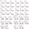
Age‐dependent immune cell dynamics and baseline hematological profiles correlating with survival outcomes. (a–h) Temporal analysis of peripheral blood immune cells in young and aged mice after injection with GA (gray), CoV1 (blue), or CoV2 (red) ssRNAs. Blood samples were collected at baseline (0‐h, n = 12–14), 4 h, 2 days, and 7 days post‐injection (n = 3–5). Analyzed parameters: (a) White blood cells (WBC); (b) Lymphocytes; (c) CD11b+ cells; (d) Natural killer (NK) cells; (e) B cells; (f) Total T cells; (g) CD4+ T cells; (h) CD8+ T cells. (i) Comparison of baseline hematological parameters among young mice, surviving (S) and non‐surviving (NS) aged mice following ssRNA injections. Parameters include WBC, lymphocytes, monocytes, neutrophils, eosinophils, and basophils. Data are presented as mean ± SD. Statistical significance was determined using two‐way ANOVA with Tukey's post hoc test for (a–h), and one‐way ANOVA with Tukey's post hoc test for (i). *p < 0.05, **p < 0.01, ***p < 0.001.
FIGURE 4
FIGURE 4
Age‐dependent cytokine dynamics, baseline profiles, and CD11b+ cell responses to SARS‐CoV ssRNAs. (a–h) Temporal changes in serum cytokine levels following injection of GA ssRNA (gray), CoV1 ssRNA (blue), or CoV2 ssRNA (red) in young surviving mice, old surviving mice, and old non‐surviving mice. Blood samples were collected at 0 (n = 10), 4 h (n = 3–7), 2 days (n = 3–5), and 7 days (n = 3–5) post‐injection. Analyzed cytokines: (a) TNF‐α; (b) IL‐6; (c) IL‐10; (d) IL‐12; (e) IFN‐α; (f) IFN‐β; (g) IFN‐γ; (h) MCP‐1. (i) Baseline (0 h) cytokine levels in young surviving (YS), old surviving (OS), and old non‐surviving (ONS) groups prior to ssRNAs challenge. See also Figure S6 for a consolidated comparison of cytokine responses across different groups at the 4‐h timepoint. (j) Cytokine secretion by CD11b+ cells isolated from young and aged mice after 24‐h in vitro exposure to various ssRNAs. Data are presented as mean ± SD. Statistical significance was determined using two‐way ANOVA with Tukey's post hoc test for (a–h), and one‐way ANOVA with Tukey's post hoc test for (i, j); *p < 0.05, **p < 0.01, ***p < 0.001.
FIGURE 5
FIGURE 5
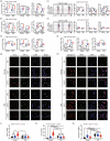
Age‐associated dysregulation of IRF7 signaling and SNARE‐mediated cytokine trafficking in CD11b+ cells. (a, b) Relative mRNA expression levels of TLR7 signaling components and downstream signaling molecules in CD11b+ cells isolated from young and aged mice after 24‐h exposure to various ssRNAs. (a) Analyzed genes in the TLR7 pathway: TLR7, MyD88, UNC93B1. (b) Analyzed genes downstream of TLR7: IRF7, p65 (NF‐κB subunit), AP1. Flow cytometric analysis of (c) phosphorylated IRF7 (p‐IRF7), total IRF7 (T‐IRF7), and p‐IRF7/T‐IRF7 ratios, as well as (d) phosphorylated p65 (p‐p65), total p65 (T‐p65), and p‐p65/T‐p65 ratios in CD11b+ cells from young and aged mice after 24‐h ssRNA treatment. Quantification of mean fluorescence intensity (MFI) of phosphorylated and total proteins were shown. (e, f) Relative mRNA expression levels of SNARE family genes in CD11b+ cells isolated from young and aged mice after 24‐h ssRNA exposure. Analyzed genes: (e) Vamp3, Vamp8, Vti1b; (f) Syntaxin‐4, Syntaxin‐6, Syntaxin‐7. (g, h) Immunofluorescence staining of CD11b+ cells from young and aged mice treated with GA, CoV1, or CoV2 ssRNAs for 24 h. (g) Co‐staining of Vti1b (red), TNF‐α (green), and DAPI (blue). (h) Co‐staining of Syntaxin‐6 (red), TNF‐α (green), and DAPI (blue). (i–k) Quantification of immunofluorescence staining showing the mean fluorescence intensity (MFI) of: (i) TNF‐α; (j) Vti1b; (k) Syntaxin‐6. Data are presented as mean ± SD. n = 5–6 per group. Statistical significance was determined using two‐way ANOVA with Tukey's post hoc test. *p < 0.05, **p < 0.01, ***p < 0.001.
FIGURE 6
FIGURE 6
Adoptive transfer of young CD11b+ cells improves survival rates and immune function in aged mice exposed to SARS‐CoV‐2 ssRNA. (a) Schematic representation of the experimental design illustrating the adoptive transfer protocol of young (Y‐Tx) and old CD11b+ cells (O‐Tx) into aged mice following CoV2 ssRNA challenge. (b) Kaplan–Meier survival analysis comparing aged mice exposed to CoV2 ssRNA with and without the adoptive transfer of CD11b+ cells. GA as a control. Statistical significance was determined using the log‐rank Mantel‐Cox test. (c) Representative lung histology (H&E staining) from aged mice and treatment (Tx) groups 7 days post‐ssRNA administration. (d) Changes in plasma cytokine levels in GA, aged survivor (S) and non‐survivor (NS), and Tx groups after 4 h of CoV2 ssRNA exposure. (e) Hematological parameters in treated mice 7 days after CoV2 ssRNA injection. Data are presented as mean ± SD. Statistical significance was determined using one‐way ANOVA with Tukey's post hoc test; p < 0.05, **p < 0.01, ***p < 0.001 compared to relevant controls. Scale bar: 100 μm.

References

    1. Ajoolabady, A. , Pratico D., Tang D., Zhou S., Franceschi C., and Ren J.. 2024. “Immunosenescence and Inflammaging: Mechanisms and Role in Diseases.” Ageing Research Reviews 101: 102540. 10.1016/j.arr.2024.102540. - DOI - PubMed
    1. Alter, G. , Suscovich T. J., Teigen N., et al. 2007. “Single‐Stranded RNA Derived From HIV‐1 Serves as a Potent Activator of NK Cells.” Journal of Immunology 178, no. 12: 7658–7666. 10.4049/jimmunol.178.12.7658. - DOI - PubMed
    1. Bartleson, J. M. , Radenkovic D., Covarrubias A. J., Furman D., Winer D. A., and Verdin E.. 2021. “SARS‐CoV‐2, COVID‐19 and the Ageing Immune System.” Nature Aging 1, no. 9: 769–782. 10.1038/s43587-021-00114-7. - DOI - PMC - PubMed
    1. Bonam, S. R. , Hazell N. C., Mathew M. J., et al. 2024. “Innate and Adaptive Immune Parameters Following mRNA Vaccination in Mice.” Vaccine 12, no. 5: 543. https://www.mdpi.com/2076‐393X/12/5/543. - PMC - PubMed
    1. Castilla, J. , Guevara M., Miqueleiz A., et al. 2021. “Risk Factors of Infection, Hospitalization and Death From SARS‐CoV‐2: A Population‐Based Cohort Study.” Journal of Clinical Medicine 10, no. 12: 2608. 10.3390/jcm10122608. - DOI - PMC - PubMed