. 2025 May 6;5(5):100821.
doi: 10.1016/j.xops.2025.100821. eCollection 2025 Sep-Oct.

Genome-Wide Association Study of Intraocular Pressure in Population-Based Cohorts in Japan: The Tohoku Medical Megabank Organization Eye Study

Affiliations

Genome-Wide Association Study of Intraocular Pressure in Population-Based Cohorts in Japan: The Tohoku Medical Megabank Organization Eye Study

Nobuo Fuse et al. Ophthalmol Sci. .

Abstract

Purpose: This study was conducted to elucidate the distribution and determinants of ocular biometric parameters and to assess the association between intraocular pressure (IOP) and single nucleotide polymorphisms (SNPs) in the Japanese population-based genome cohort studies.

Design: Cross-sectional analysis involving genome-wide association studies (GWASs).

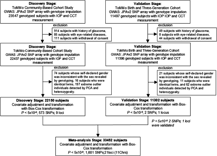
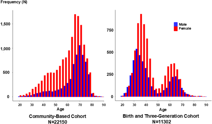
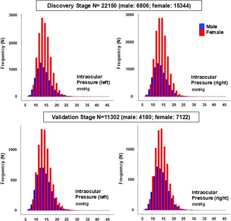
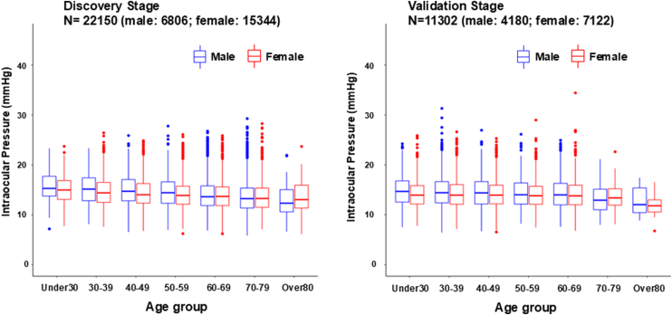
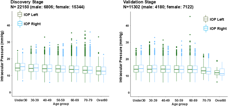
Participants: In total, 22 150 participants aged >18 years from the population cohort (Community-Based Cohort [CommCohort]) and 11 302 participants from the Birth and Three-Generation (BirThree) Cohort of the Tohoku Medical Megabank Organization Eye Study were examined.

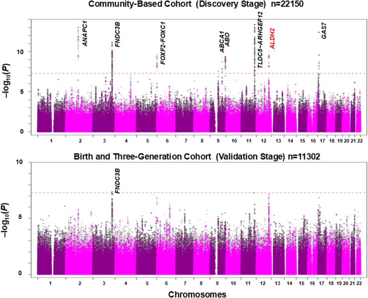
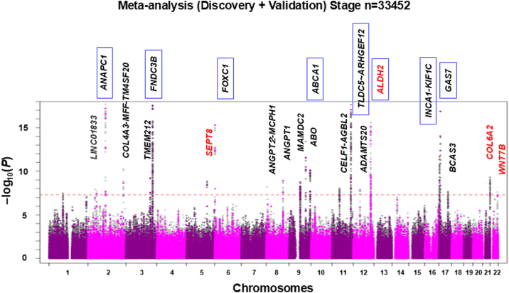
Methods: Participant underwent interviews, ophthalmic and physiological examinations, laboratory tests, and microarray analyses. Genome-wide association studies were conducted in the CommCohort (discovery stage) and the BirThree Cohort (replication stage), followed by a meta-analysis. Associations of SNPs and IOP were evaluated using a genome-wide significance threshold (5 × 10- 8).

Main outcome measures: Association of SNPs with IOP and distributions of IOP by sex and age.

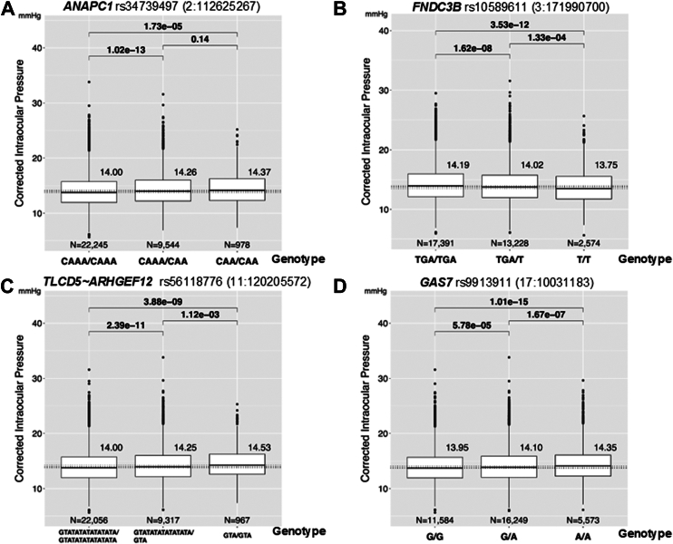
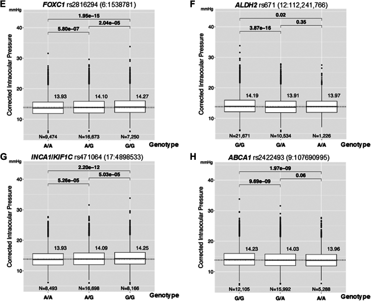
Results: In the discovery stage, the mean IOP of the right and left eye was 13.95 and 14.02 mmHg, respectively. In the replication stage, the corresponding values were 14.32 and 14.27 mmHg, respectively. A significant age-related reduction in IOP was observed in both stages (P < 0.001). Genome-wide association studies identified 573 and 2 genome-wide significant SNPs in the discovery and replication stages, respectively. Meta-analysis revealed 1601 significant SNPs across 21 loci on 11 chromosomes (Chrs). Of these loci, 17 were previously known to be associated with IOP or glaucoma, while four-septin-8 (SEPT8; Chr5), aldehyde dehydrogenase 2 (ALDH2; Chr12), collagen type VI alpha 2 chain (COL6A2; Chr21), and Wnt family member 7B (WNT7B; Chr22)-were newly identified.

Conclusions: This large-scale GWAS in a Japanese population identified 21 loci associated with IOP, including 4 novel loci. The findings highlight both genetic similarities and population-specific variations in SNPs influencing IOP and provide valuable insights to enhance eye health care, including glaucoma management.

Financial disclosures: Proprietary or commercial disclosure may be found in the Footnotes and Disclosures at the end of this article.

Keywords: Genome-wide association study (GWAS); Intraocular pressure; Population-based cohort study.


Figures

Figure 1
Figure 1
Genome-wide association study design for investigating genetic correlates of IOP across 2 cohorts. The inclusion criteria for the 2 population cohorts and the overall scheme of GWAS for the discovery and validation stages are presented. The discovery-stage GWAS utilized the CommCohort Study, comprising 22 150 genotyped participants with IOP measurements. The validation-stage GWAS was conducted using the BirThree Cohort Study, which included 11 302 genotyped participants with IOP measurements, after applying the exclusion criteria. Associations between SNPs and IOP were assessed using a genome-wide significance threshold (P < 5 × 10⁸) in each stage and in a combined meta-analysis involving a total of 33 452 participants. BirThree = Birth and Three-Generation; CCT = central corneal thickness; CommCohort = Community-Based Cohort; GWAS = genome-wide association study; IOP = intraocular pressure; PCA = principal component analysis; SNP = single nucleotide polymorphism.
Figure 2
Figure 2
Age distribution of the study population. Intraocular pressure examinations were conducted at 7 community support centers. For the discovery stage, a total of 22 150 participants were recruited (mean age 61.76 ± 12.40 years; 6806 men and 15 344 women). For the validation stage, 11 302 participants were included (mean age 43.73 ± 13.48 years; 4180 men and 7122 women). In the discovery stage, the majority of participants were in their 60s to 70s, whereas the validation stage exhibited 2 age peaks, primarily in the 30s and 60s.
Figure 3
Figure 3
Intraocular pressure distribution across age groups. The mean IOP was 13.95 mmHg (95% CI: 13.91–13.99) in the right eye and 14.32 mmHg (95% CI: 14.27–14.38) in the left eye during the discovery stage. In the validation stage, the mean IOP was 14.02 mmHg (95% CI: 13.98–14.06) in the right eye and 14.27 mmHg (95% CI: 14.21–14.33) in the left eye. Notably, IOP in both the right and left eyes exhibited significant age-related variations among both female and male participants. CI = confidence interval; IOP = intraocular pressure.
Figure 4
Figure 4
Age-related decrease in IOP values showed a significant decline with advancing age in both men and women (P < 0.001) during the discovery stage (left panel). This age-associated reduction in IOP was consistently reproduced in the validation-stage data (right panel). IOP = intraocular pressure.
Figure 5
Figure 5
Age-dependent right–left differences in IOP. The most pronounced differences in IOP between the right and left eyes were observed among younger participants. During the discovery stage, the right eye exhibited significantly higher IOP than the left eye in individuals in their 30s (P < 0.035, left panel). Similarly, in the validation stage, the right eye showed significantly higher IOP compared to the left eye in participants under 30 (P = 0.009) and in their 30s (P = 0.007, right panel). This trend persisted in participants under 50 years of age in both stages. However, the relationship reversed in older age groups, with the left eye demonstrating higher IOP values in elderly participants. IOP = intraocular pressure.
Figure 6
Figure 6
Background characteristics associated with IOP. The associations between IOP values and participants' background characteristics were analyzed. Significant correlations were observed between IOP and several factors, including central corneal thickness, serum glucose levels, total cholesterol levels, platelet count, lung function (percent-predicted forced expiratory volume in the first second), as well as systolic and diastolic blood pressures (data from both discovery stages are presented). IOP = intraocular pressure.
Figure 7
Figure 7
Genome-wide association study of IOP in the discovery and validation stage. The results of the GWAS for IOP conducted in the discovery and validation stages are presented. Data for both directly genotyped and imputed SNPs are visualized in the Manhattan plot. The y-axis depicts –log10(P) values for associations with IOP, while the x-axis represents chromosomes and base-pair positions based on human genome build 37. The horizontal red dotted line denotes the genome-wide significance threshold (P < 5.0 × 10⁸). Previously reported genes (loci) are labeled in black, while newly identified genes (locus) from this study are highlighted in red. ABO = alpha 1-3-N-acetylgalactosaminyltransferase and alpha 1-3-galactosyltransferase; ALDH2 = aldehyde dehydrogenase 2; ANAPC1 = anaphase promoting complex subunit 1; FNDC3B = fibronectin type III domain-containing protein 3B; FOXF2 = forkhead box F2; GAS7 = growth arrest specific 7; GWAS = genome-wide association study; IOP = intraocular pressure; SNP = single nucleotide polymorphism.
Figure 8
Figure 8
Genome-wide association study of intraocular pressure in the meta-analysis. The results of the GWAS for intraocular pressure in the meta-analysis are presented. Data for both directly genotyped and imputed SNPs are visualized in the Manhattan plot, with details consistent with those in Figure 7. Previously reported genes (loci) are labeled in black, while newly identified genes (loci) from this study are highlighted in red. Closed boxes indicate the top 8 SNPs. ANAPC1 = anaphase promoting complex subunit 1; ANGPT2 = angiopoietin 2; ANGPT1 = angiopoietin 1; ABO = alpha 1-3-N-acetylgalactosaminyltransferase and alpha 1-3-galactosyltransferase; ABCA1 = ATP binding cassette subfamily A member 1; AGBLG2 = AGBL carboxypeptidase 2; ADAMTS20 = ADAM metallopeptidase with thrombospondin type 1 motif 20; ALDH2 = aldehyde dehydrogenase 2; BCAS3 = BCAS3 microtubule associated cell migration factor; COL4A3 = collagen type IV alpha 3 chain; ARHGEF12 = Rho Guanine Nucleotide Exchange Factor 12; CELF1 = CUGBP Elav-like family member 1; COL6A2 = collagen type VI alpha 2 chain; FNDC3B = fibronectin type III domain-containing protein 3B; FOXC1 = forkhead box C1; GWAS = genome-wide association study; GAS7 = growth arrest specific 7; INCA1 = inhibitor of CDK, cyclin A1 interacting protein 1; KIF1C = kinesin family member 1C; LINC01833 = long intergenic non-protein coding RNA 1833; MFF = mitochondrial fission factor; MCPH1 = microcephalin 1; MAMDC2 = MAM domain containing 2; SNP = single nucleotide polymorphism; SEPT8 = septin-8; TM4SF20 = transmembrane 4 L six family member 20; TMEM212 = transmembrane protein 212; TLDC5 = TLC domain containing 5; WNT7B = Wnt family member 7B.
Figure 9
Figure 9
Effects of validated SNPs on the phenotypic values of IOP. The boxplot illustrates the effects of genotypes for the validated top 8 SNPs on IOP values. A,ANAPC1 rs34739497, B,FNDC3B rs10589611, C,TLCD5ARHGEF12 rs56118776, D,GAS7 rs9913911, E,FOXC1 rs2816294, F,ALDH2 rs671, G,INCA1/KIF1C rs471064, and H,ABCA1 rs2422493. All heterozygous variants exhibit measurable effects, categorized into 2 types: those associated with an increase in IOP (ANAPC1, TLCD5∼ARHGEF12, GAS7, FOXC1, and INCA1/KIF1C) and those associated with a decrease in IOP (FNDC3B, ANAPC1, ALDH2, and ABCA1). ANAPC1 = anaphase promoting complex subunit 1; FNDC3B = fibronectin type III domain-containing protein 3B; GAS7 = growth arrest specific 7; IOP = intraocular pressure; SNP = single nucleotide polymorphism.
Figure 9
Figure 9
Effects of validated SNPs on the phenotypic values of IOP. The boxplot illustrates the effects of genotypes for the validated top 8 SNPs on IOP values. A,ANAPC1 rs34739497, B,FNDC3B rs10589611, C,TLCD5ARHGEF12 rs56118776, D,GAS7 rs9913911, E,FOXC1 rs2816294, F,ALDH2 rs671, G,INCA1/KIF1C rs471064, and H,ABCA1 rs2422493. All heterozygous variants exhibit measurable effects, categorized into 2 types: those associated with an increase in IOP (ANAPC1, TLCD5∼ARHGEF12, GAS7, FOXC1, and INCA1/KIF1C) and those associated with a decrease in IOP (FNDC3B, ANAPC1, ALDH2, and ABCA1). ANAPC1 = anaphase promoting complex subunit 1; FNDC3B = fibronectin type III domain-containing protein 3B; GAS7 = growth arrest specific 7; IOP = intraocular pressure; SNP = single nucleotide polymorphism.

Similar articles

References

    1. Quigley H.A., Broman A.T. The number of people with glaucoma Worldwide in 2010 and 2020. Br J Ophthalmol. 2006;90:262–267. - PMC - PubMed
    1. Jonas J.B., Aung T., Bourne R.R., et al. Glaucoma. Lancet. 2017;390:2183–2193. - PubMed
    1. Heijl A., Leske M.C., Bengtsson B., et al. Reduction of intraocular pressure and glaucoma progression: results from the Early Manifest Glaucoma Trial. Arch Ophthalmol. 2002;120:1268–1279. - PubMed
    1. Leske M.C., Heijl A., Hyman L., et al. Predictors of long-term progression in the early manifest glaucoma trial. Ophthalmology. 2007;114:1965–1972. - PubMed
    1. The effectiveness of intraocular pressure reduction in the treatment of normal-tension glaucoma. Collaborative Normal-Tension Glaucoma Study Group. Am J Ophthalmol. 1998;126:498–505. - PubMed

LinkOut - more resources