. 2025 Jun 23:20:8023-8039.
doi: 10.2147/IJN.S519906. eCollection 2025.

Developing the Strategy to Use Silk Spheres for Efficient, Targeted Delivery of Oligonucleotide Therapeutics to Cancer Cells

Affiliations

Developing the Strategy to Use Silk Spheres for Efficient, Targeted Delivery of Oligonucleotide Therapeutics to Cancer Cells

Sara Molenda et al. Int J Nanomedicine. .

Abstract

Introduction: Oligonucleotide-based drugs, such as siRNA, hold great promise for disease treatment, including cancer. However, their clinical application has challenges related to cell-specific delivery and susceptibility to degradation. The use of drug delivery systems (DDS) may address these problems. Nanoparticles of bioengineered spider silk demonstrate significant potential as DDS due to their biocompatibility and biodegradability. Another advantage of this material is the possibility of functionalization, which allows the control of its property. The main objective of this study was to develop a strategy for targeted delivery of oligonucleotide-based therapeutics into cancer cells using bioengineered silk technology.

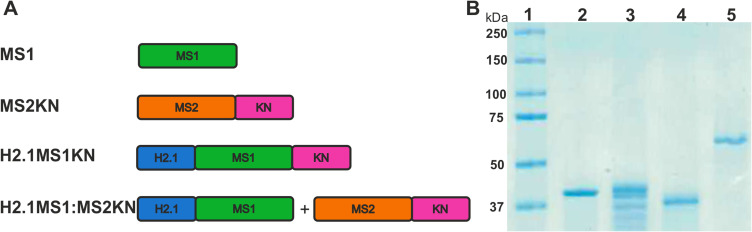
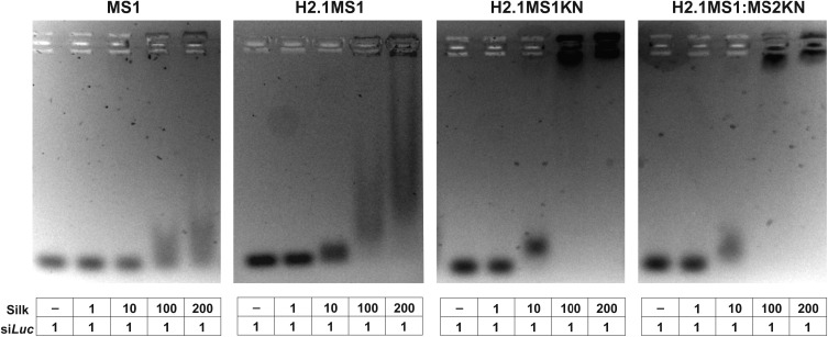
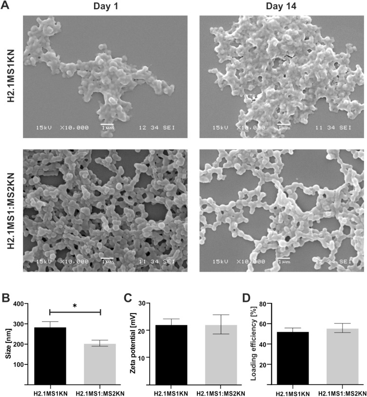
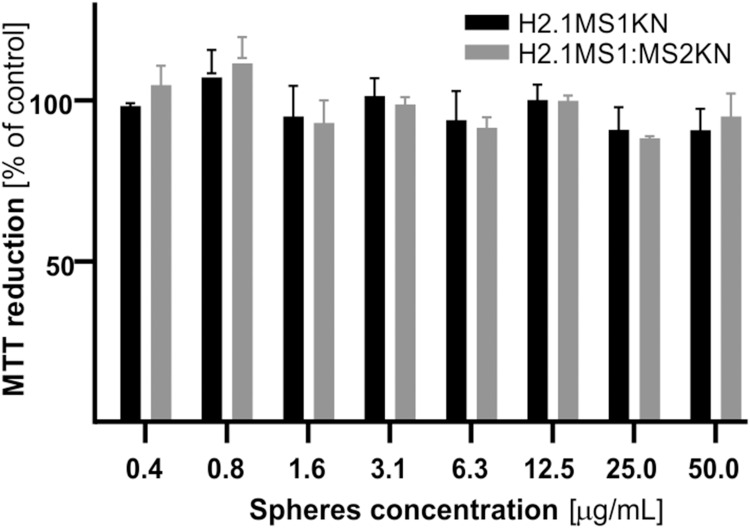
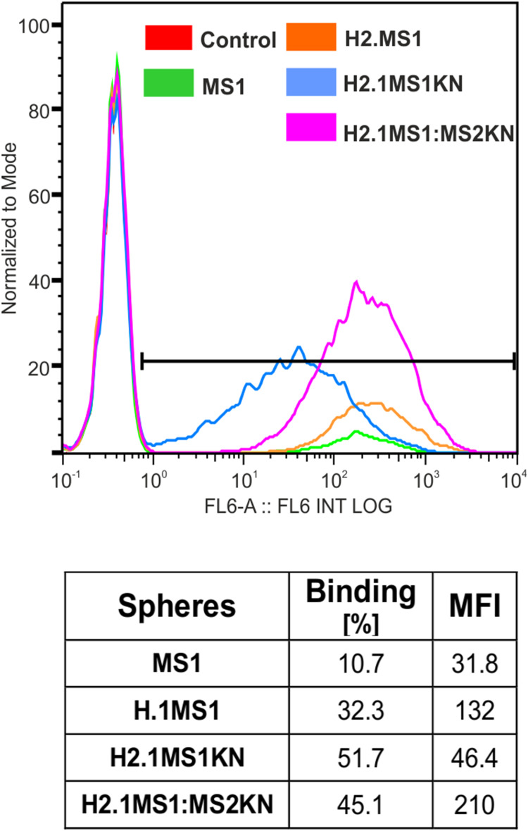
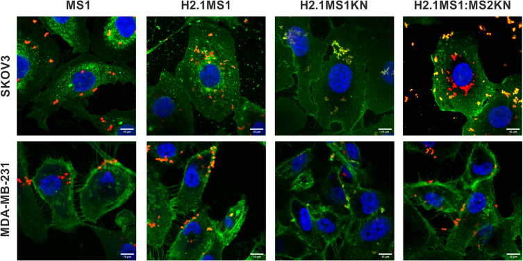
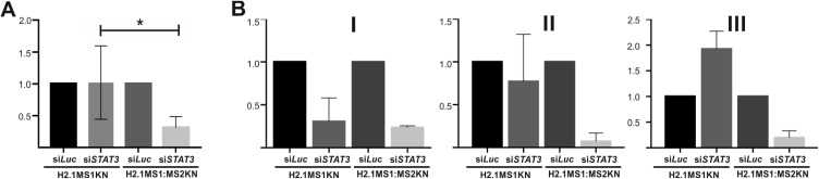
Materials and methods: Two spider silk spheres that bind oligonucleotides and target cancer cells that overexpress HER2 (HER2+) were constructed. One type of sphere was made of a newly designed silk, H2.1MS1KN, which contained two functional peptides: H2.1 for binding HER2 and KN for binding oligonucleotide. The second type of sphere was formed of a blend of two previously described proteins, H2.1MS1 and MS2KN; these proteins differed not only in the functional domain (H2.1 vs KN) but also in the sequence of silk (MS1 vs MS2). The ability of proteins to bind oligonucleotides was analyzed via gel electrophoresis. The biophysicochemical properties of particles were analyzed using an SEM, NanoSight, ZetaSizer, flow cytometry, and scanning confocal microscopy. The silk particle potential was analyzed using siRNA for silencing STAT3 expression in the HER2+ breast cancer model.

Results: Both H2.1MS1KN and H2.1MS1:MS2KN proteins efficiently bound nucleic acid. H2.1MS1:MS2KN formed smaller spheres than H2.1MS1KN. Although both H2.1MS1KN and blended H2.1MS1:MS2KN spheres were effectively loaded with oligonucleotides, only H2.1MS1:MS2KN spheres delivered siRNA to HER2+ cancer cells that successfully silenced STAT3 expression.

Conclusion: Not only the selection of functional peptides but also their quantity and type of silk is crucial when developing an effective silk-based DDS for delivering active siRNA.

Keywords: STAT3; bioengineered spider silk; cancer therapy; siRNA-delivery; spheres; targeted delivery.


Conflict of interest statement

The authors report no conflicts of interest in this work.

Figures

Figure 1
Figure 1
Spider silks constructs and SDS-PAGE analysis of silks. (A) A schematic representation of the spider silk protein constructs. Building blocks: - MS1 – MS1 silk (green), MS2 – MS2 silk (Orange), KN – nucleic acid binding peptide (pink), H2.1 – HER2 binding peptide (blue). Constructs: MS1 – control MS1 silk, MS2KN – MS2 silk functionalized with KN peptide, H2.1MS1KN - MS1 silk double-functionalized with H2.1 and KN peptides, H2.1MS1:MS2KN – blend of H2.1MS1 (MS1 silk functionalized with H2.1 peptide) and MS2KN (MS2 silk functionalized with KN peptide) silks. (B) Electrophoretic analysis of control and hybrid proteins using 12.5% SDS-PAGE gel. 1. Protein molecular weight marker (Precision Plus Protein™ Kaleidoscope™ Prestained Protein Standards, BioRad), 2. MS1 silk (MW 39.25 kDa), 3. H2.1MS1 silk (MW 41,68 kDa), 4. H2.1MS1KN silk (MW 38.56 kDa), and 5. MS2KN silk (MW 48,83 kDa).
Figure 2
Figure 2
Efficiency of siRNA binding to spider silk proteins. The siLuc and the soluble bioengineered silks (MS1 (control), H2.1MS1 (control), H2.1MS1KN, and blend of H2.1MS1 and MS2KN (H2.1MS1:MS2KN) were mixed at different molecular ratios and incubated for 5 min at room temperature. The binding of oligonucleotides to silk proteins was analyzed using agarose gel electrophoresis.
Figure 3
Figure 3
Physicochemical properties of the H2.1MS1KN and H2.1MS1:MS2KN spheres. (A) The morphology of H2.1MS1KN and H2.1MS1:MS2KN spheres analyzed by SEM (scale bar - 1 μm). (B) The size of H2.1MS1KN and H2.1MS1:MS2KN spheres (D90) was measured using NanoSight at the spheres concentration of 0.05 mg/mL (C) Zeta potential of H2.1MS1KN and H2.1MS1:MS2KN spheres was measured at 25 °C in ddH2O at the sphere concentration of 0.01 mg/mL. (D) Loading efficiency of iFam-siScr into silk spheres. The results are expressed as the mean ± SD. * Indicates significance at p ≤ 0.05.
Figure 4
Figure 4
Cytotoxicity evaluation of silk spheres. SKOV3 ovary cancer cells were incubated with various concentrations of H2.1MS1KN and H2.1MS1:MS2KN silk spheres, and cell metabolic activity was assessed using the MTT assay. The percentage of MTT reduction was determined relative to untreated control cells. Data are presented as the mean of three independent experiments, with error bars representing the standard error of the mean.
Figure 5
Figure 5
Flow cytometry analysis of sphere binding by HER+ cancer cells. The human SKOV3 ovary cancer cells were incubated for 1 h with Cy5-labeled H2.1MS1KN or H2.1MS1:MS2KN silk spheres and analyzed by flow cytometry.
Figure 6
Figure 6
Confocal microscopy analysis of the cellular uptake of H2.1MS1KN, H2.1MS1:MS2KN and MS1 control silk spheres. The HER2-overexpressing human ovary cancer cells SKOV3, and HER2-negative human breast cancer cells MDA-MB-231 were incubated with silk particles for 4 h. FITC-Concanavalin A was used for staining cell surface carbohydrates (green), silk spheres were labeled with Cy5 (red), and nucleus was stained with DAPI (blue). Scale bar - 10 μm.
Figure 7
Figure 7
Analysis of STAT3 mRNA level after treatment with siSTAT3 delivered by H2.1MS1KN and H2.1MS1:MS2KN spheres. (A) The HER2+ SKOV3 cancer cells were incubated for 48 h with H2.1MS1KN or H2.1MS1MS2KN spheres loaded with siSTAT3 (250nM) or siLuc(250 nM) and then qPCR analysis was performed. STAT3 mRNA expression level was normalized to GAPDH expression. *Indicates significance at p ≤ 0.05. Results are expressed as means of six independent experiments and error bars show the standard error of the mean. (B) After being loaded with siRNA, the spheres were stored for (I) 1 week, II) 2 weeks, or III) 3 weeks and then used for STAT3 silencing. Each experiment was performed once after indicated time of storage (I–III) in technical repetitions (mean ± SD). The mean value and the standard error of the mean. of the silencing effect of siSTAT3 delivered by H2.1MS1KN shown in 7A were calculated based on the results obtained for stored spheres from different time points.

Similar articles

References

    1. Kaur R, Bhardwaj A, Gupta S. Cancer treatment therapies: traditional to modern approaches to combat cancers. Mol Biol Rep. 2023;50(11):9663–9676. doi: 10.1007/s11033-023-08809-3 - DOI - PubMed
    1. Cesur‐Ergün B, Demir‐Dora D. Gene therapy in cancer. J Gene Med. 2023;25(11):e3550. doi: 10.1002/jgm.3550 - DOI - PubMed
    1. Chung SS, Giehl N, Wu Y, Vadgama JV. STAT3 activation in HER2-overexpressing breast cancer promotes epithelial-mesenchymal transition and cancer stem cell traits. Int J Oncol. 2014;44(2):403–411. doi: 10.3892/ijo.2013.2195 - DOI - PMC - PubMed
    1. Molenda S, Sikorska A, Florczak A, Lorenc P, Dams-Kozlowska H. Oligonucleotide-based therapeutics for STAT3 targeting in cancer—drug carriers matter. Cancers. 2023;15(23):5647. doi: 10.3390/cancers15235647 - DOI - PMC - PubMed
    1. Hossain DMS, Dos Santos C, Zhang Q, et al. Leukemia cell–targeted STAT3 silencing and TLR9 triggering generate systemic antitumor immunity. Blood. 2014;123(1):15–25. doi: 10.1182/blood-2013-07-517987 - DOI - PMC - PubMed

MeSH terms

LinkOut - more resources