

This is a preprint.
Bridging Neuroimaging and Neuropathology: A Comprehensive Workflow for Targeted Sampling of White Matter Lesions
- PMID: 40585178
- PMCID: PMC12204417
- DOI: 10.1101/2025.06.08.25329217
Bridging Neuroimaging and Neuropathology: A Comprehensive Workflow for Targeted Sampling of White Matter Lesions
Update in
-
Bridging Neuroimaging and Neuropathology: A Comprehensive Workflow for Targeted Sampling of White Matter Lesions.J Neuroimaging. 2025 Sep-Oct;35(5):e70094. doi: 10.1111/jon.70094. J Neuroimaging. 2025. PMID: 41126483 Free PMC article. Review.
Abstract
Background and purpose: White matter lesions are common imaging biomarkers associated with aging and neurodegenerative diseases, yet their underlying pathology remains unclear due to limitations in imaging-based characterization. We aim to develop and validate a comprehensive workflow enabling precise MRI-guided histological sampling of white matter lesions to bridge neuroimaging and neuropathology.
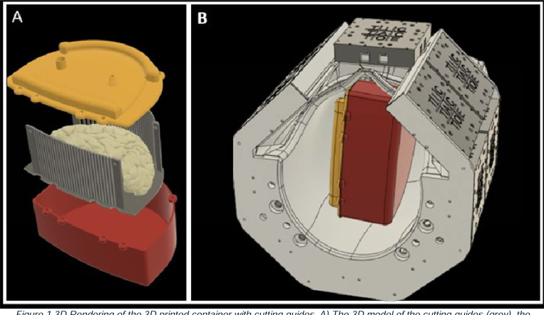
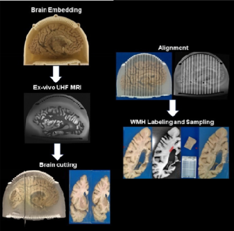
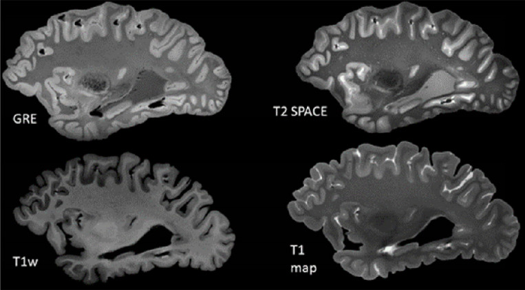
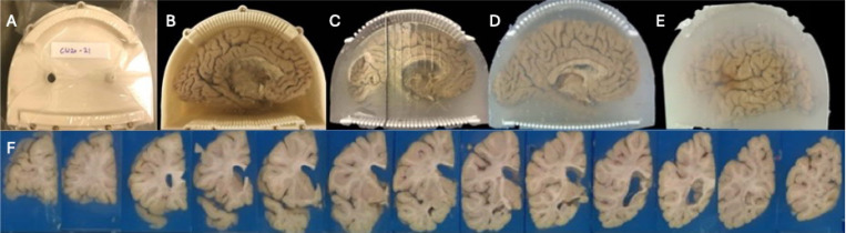
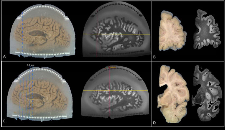
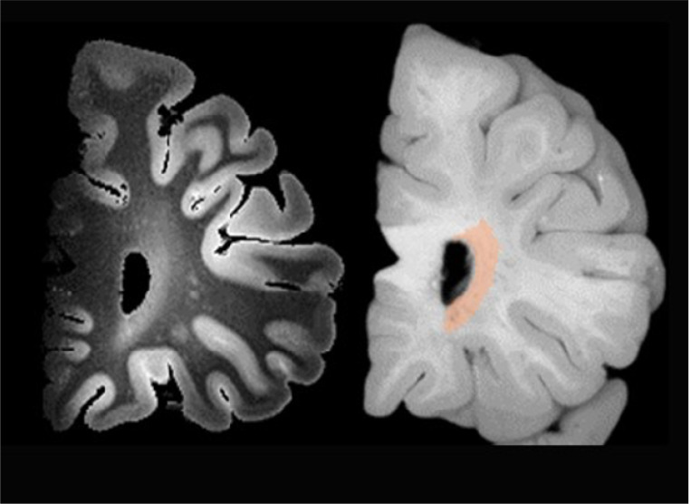
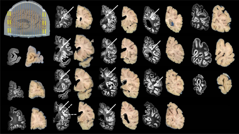
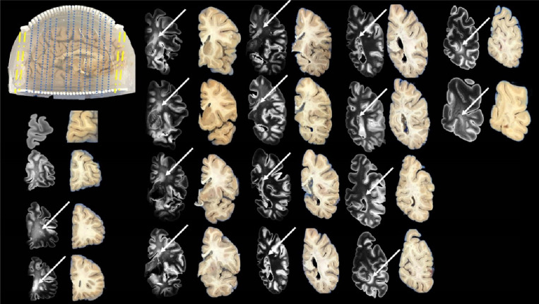
Methods: We establish a workflow integrating agarose-saccharose brain embedding, ultra-high field 7T MRI acquisition, reusable 3D-printed cutting guides, and semi-automated MRI-blockface alignment. Postmortem brains are stabilized in the embedding medium and scanned using optimized MRI protocols. Coronal sectioning is guided by standardized 3D-printed cutting guides, and knife traces are digitally matched to MRI planes. White matter lesions are segmented on MRI and aligned for histopathological sampling. This approach is validated in over 100 postmortem human brains.
Results: The workflow enables reproducible brain sectioning, minimizes imaging artifacts, and achieves precise spatial alignment between MRI and histology. Consistent, high-resolution MRI data facilitated accurate lesion detection and sampling. The use of standardized cutting guides and alignment protocols reduce variability and improve efficiency.
Conclusions: Our cost-effective, scalable workflow reliably links neuroimaging findings with histological analysis, enhancing the understanding of white matter lesion pathology. This framework holds significant potential for advancing translational research in aging and neurodegenerative diseases.
Keywords: 3D printing; MRI-guided histology; White matter lesions; neuroimaging workflow; postmortem brain imaging; ultra-high field MRI.
Conflict of interest statement
The authors declare no commercial or financial conflicts of interest relevant to this study.
Figures
References
-
- Hajnal J.V., et al. , Use of Fluid Attenuated Inversion Recovery (FLAIR) Pulse Sequences in MRI of the Brain. Journal of Computer Assisted Tomography, 1992. 16(6): p. 841–844. - PubMed
-
- Sachdev P.S., et al. , Is Alzheimer’s a disease of the white matter? Current Opinion in Psychiatry, 2013. 26(3): p. 244–251. - PubMed
Publication types
Grants and funding
LinkOut - more resources
Full Text Sources