. 2025 Jun 19;7(2):lqaf084.
doi: 10.1093/nargab/lqaf084. eCollection 2025 Jun.

Differential cellular communication inference framework for large-scale single-cell RNA-sequencing data

Affiliations

Differential cellular communication inference framework for large-scale single-cell RNA-sequencing data

Giulia Cesaro et al. NAR Genom Bioinform. .

Abstract

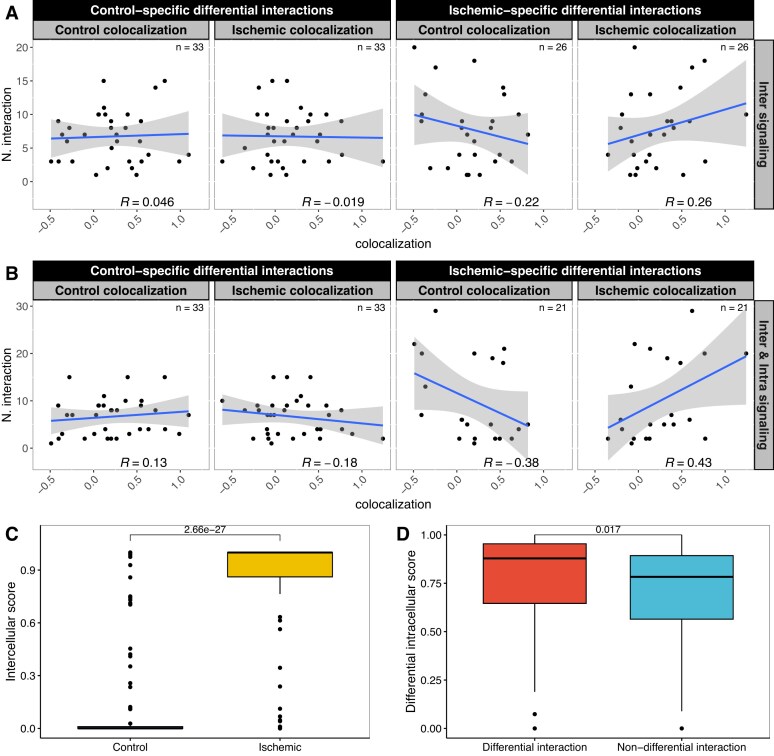
Single-cell transcriptomics data have been widely used to characterize biological systems, particularly in studying cell-cell communication, which plays a significant role in many biological processes. Despite the availability of various computational tools for inferring cellular communication, quantifying variations across different experimental conditions at both intercellular and intracellular levels remains challenging. Moreover, available methods are in general limited in terms of flexibility in analyzing different experimental designs and the ability to visualize results in an easily interpretable way. Here, we present a generalizable computational framework designed to infer and support differential cellular communication analysis across two experimental conditions from large-scale single-cell transcriptomics data. The scSeqCommDiff tool employs a statistical and network-based computational approach for characterizing altered cellular cross-talk in a fast and memory-efficient way. The framework is complemented with CClens, a user-friendly Shiny app to facilitate interactive analysis of inferred cell-cell communication. Validation through spatial transcriptomics data, comparison with other tools, and application to large-scale datasets (including a cell atlas) confirms the reliability, scalability, and efficiency of the framework. Moreover, the application to a single-nucleus transcriptomics dataset shows the validity and ability of the proposed workflow to support and unravel alterations in cell-cell interactions among patients with amyotrophic lateral sclerosis and healthy subjects.


Conflict of interest statement

None declared.

Figures

Graphical Abstract
Graphical Abstract
Figure 1.
Figure 1.
Schematic overview of the proposed two-step framework for analyzing differential cellular communication. The framework requires as input both a priori biological knowledge of cell–cell communication (built-in or user-specified resources) and a normalized scRNA-seq gene expression matrix (in a conditions-aware or multi-sample conditions-aware setting). As a first step, the scSeqCommDiff tool identifies and quantifies alterations in cellular communication across experimental conditions (A versus B) at both intercellular and intracellular levels. Differential intercellular communication is assessed by examining the strength of ligand–receptor interaction through different built-in scoring schemes and using statistical methods to determine significance. For intracellular signaling, scSeqCommDiff incorporates network analysis and statistical tests to identify differential transcriptional regulation within downstream signaling pathways. The implementation is optimized for fast and memory-efficient analysis using shared memory or sparse matrix data representations and parallel computations in order to handle large-scale datasets efficiently. As a second step, the CClens visualization tool supports the interpretation and visualization of scSeqCommDiff outputs, as well as output from other cell–cell communication inference tools. CClens offers flexible data filtering criteria for inspection and exploration, as well as providing rich visualizations to enhance understanding and analysis of differential cellular communication. Created in BioRender. Cesaro, G. (2025) https://BioRender.com/7ji4vvb.
Figure 2.
Figure 2.
Results of agreement with spatial co-localization data. (A and B) Spatial co-localization analysis of the top prioritized differential cellular communication analysis, considering only intercellular signaling (A) or both intercellular and intracellular signaling (B). The number of prioritized differential ligand–receptor interactions (y-axis) between cell type pairs within the top n= 250 condition-specific ligand–receptor interactions is computed for control and ischemic conditions. Spatial co-localization (x-axis) of each cell type pair is computed in both control and ischemic samples. Each data point represents a distinct cell type pair involved in at least one interaction among the top-ranked differential interactions predicted by scSeqCommDiff (with the number of points indicated in the upper corner of each panel). Pearson correlation between the number of prioritized differential interactions and co-localization is shown, along with blue lines indicating linear model fits and gray bands representing 95% confidence level intervals. (C) Boxplot showing the distribution of intercellular scores (formula image) of predicted differential interactions for cell type pairs statistically co-localized in ischaemic samples compared with controls. One-sided P-value from the Wilcoxon signed-rank test is shown. (D) Boxplot representing the distribution of differential intracellular scores (formula image) associated with predicted differential and non-differential interaction for cell type pairs statistically co-localized in ischemic samples. The P-value from the one-sided Wilcoxon signed-rank test is shown.
Figure 3.
Figure 3.
Results of benchmarking using DES. The box plot illustrates the DES distributions for various tools applied to the human myocardial infarction dataset (A) and the multiple sclerosis dataset (B). Additionally, tools are categorized based on their methodological approach, distinguishing between conditions-aware (left) and multi-sample conditions-aware (right) scenarios. The tools are ranked by their median DES values.
Figure 4.
Figure 4.
Evaluation of the computational performance of scSeqCommDiff across two different datasets, i.e. the Pineda et al. dataset and LuCA atlas. (A) Multi-sample conditions-aware scenario. Maximum observed memory overhead and running time for different data structures (big.matrix and sparseMatrix objects) when applying scSeqCommDiff in a multi-sample conditions-aware setting using serial mode. (B) Conditions-aware scenario. Maximum observed memory overhead and running time over an increasing number of cores for different data structures (big.matrix and sparseMatrix objects) when applying scSeqCommDiff in a conditions-aware scenario.
Figure 5.
Figure 5.
Differential cellular communication analysis of ALS versus PN in the multi-sample conditions-aware scenario with CClens. (A) Screenshot of CClens displaying the filtering options. Results are filtered to analyze interactions that are statistically overcommunicating in ALS, with evidence of an altered intracellular response. (B) Interactive chord diagram displaying the number of interactions across cell type pairs. (C) Dot plot showing the top-ranked outgoing interactions (x-axis) across different cell type pairs (y-axis) involving astrocytes as the sender cell type. The intercellular score for each interaction is mapped as dot size, the differential intracellular score as a color fill, and the experimental condition is encoded by the colored stroke. (D) Dot plot displaying interactions involving GAD enzymes and GABA receptors (x-axis), astrocytes as the sender cell type, and microglia as the receiver cell type (y-axis). The values of intercellular score for each interaction are mapped as dot size, the differential intracellular score as colored fill, and the experimental condition is encoded by the colored stroke.

Similar articles

References

    1. Cihankaya H, Theiss C, Matschke V Significance of intercellular communication for neurodegenerative diseases. Neural Regen Res. 2022; 17:1015. - PMC - PubMed
    1. Garden GA, La Spada AR Intercellular (Mis)communication in neurodegenerative disease. Neuron. 2012; 73:886–901. 10.1016/j.neuron.2012.02.017. - DOI - PMC - PubMed
    1. Nisar S, Hashem S, Macha MA et al. Exploring dysregulated signaling pathways in cancer. Curr Pharm Des. 2020; 26:429–45. 10.2174/1381612826666200115095937. - DOI - PubMed
    1. Cesaro G, Milia M, Baruzzo G et al. MAST: a hybrid multi-agent spatio-temporal model of tumor microenvironment informed using a data-driven approach. Bioinform Adv. 2022; 2:vbac092. 10.1093/bioadv/vbac092. - DOI - PMC - PubMed
    1. Schiavone S, Contursi A, Grande R et al. Therapeutic targeting of dysregulated cellular communication. Ann Transl Med. 2017; 5:222. 10.21037/atm.2017.03.62. - DOI - PMC - PubMed

LinkOut - more resources