Review
. 2025 Jun 13:52:338-365.
doi: 10.1016/j.bioactmat.2025.06.005. eCollection 2025 Oct.

Emerging technologies towards extracellular vesicles large-scale production

Affiliations
Review

Emerging technologies towards extracellular vesicles large-scale production

Junjie Huang et al. Bioact Mater. .

Abstract

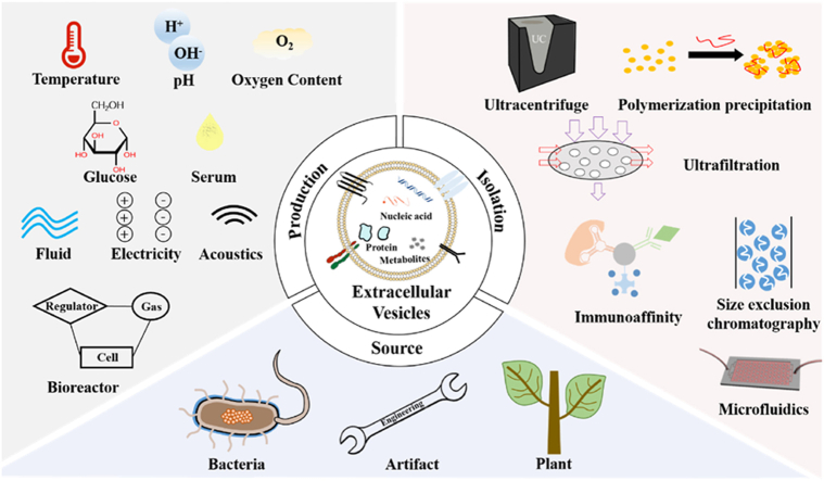
Extracellular vesicles (EVs), which carry bioactive components such as proteins and nucleic acids, reflect the physiological state of their parent cells and play a key role in mediating complex intercellular signaling. Leveraging these unique characteristics, researchers have explored their potential applications in cell therapy, non-invasive biopsies, and tissue regeneration. Therefore, standardized and scalable methods for EVs production and purification are crucial for clinical application and therapeutic settings. However, the limited yields of traditional production and isolation methods have hampered full potential of EVs. In this review, we will introduce strategies aimed at enhancing EV production include optimizing cell yield, expanding cell culture scale, and exploring alternative EVs production sources such as non-mammalian organisms and artificially produced vesicles. Various approaches as well as the bioreactors for controlling cell culture to enhance EVs production, will be introduced in detail. These approaches include regulation of culture parameters, culture medium components, and external stimuli. Additionally, the comparison between traditional ultracentrifugation methods and advance microfluidic isolating methods will be analyzed and discussed. Finally, we will introduce the potential challenges of transitioning EVs from basic research to clinical application and further discuss the future prospects. As the technology advances and different methods are integrated, there is significant potential to enable large-scale EVs production and improve their clinical translation.

Keywords: Bioreactor; Clinical translation; Extracellular vesicles; Isolation; Large-scale.


Conflict of interest statement

The authors declare the following financial interests/personal relationships which may be considered as potential competing interests: Yuanjin Zhao reports financial support was provided by National Natural Science Foundation of China. Yuanjin Zhao reports financial support was provided by National Key Research and Development Program of China. Yuanjin Zhao reports financial support was provided by Guangdong Basic and Applied Basic Research Foundation. Yuanjin Zhao reports financial support was provided by Clinical Trials from Nanjing Drum Tower Hospital. Yuanjin Zhao reports financial support was provided by Shenzhen Fundamental Research Program. Yuanjin Zhao is an editorial board member for Bioactive Materials and was not involved in the editorial review or the decision to publish this article. Other authors declare that there are no competing interests.

Figures

Image 1
Graphical abstract
Fig. 1
Fig. 1
The classical pathway of exosome origin. Reproduced with permission [59]. Copyright 2021, Elsevier.
Fig. 2
Fig. 2
(a) Schematic view of hollow fiber bioreactor. Reproduced under a Creative Commons Attribution 4.0 International License [65]. Copyright 2020, The Authors, published by Springer Nature. (b) (i) Optical microscopy image and (ii) confocal images of the stem cells loaded on 3D collagen hydrogel, green represents viable cells. Reproduced with permission [66]. Copyright 2022, Elsevier. (c) Fluorescent images of the live/dead staining for stem cells cultured on microspheres with diverse sizes and membranes for 5 days. The x-axis represents cultivation on a flat and three different-sized carriers. Reproduced with permission [67]. Copyright 2022, Elsevier. (d) Gross morphology of stem cells drop formed by (i) hanging-drop technique and (ii) poly (2-hydroxyethyl methacrylate) coating. Reproduced with permission [68]. Copyright 2018, Springer Nature. (e) Optical microscopy images of cells' spheroid morphology deal with different conditions. Cell MNP: cell and magnetic nanoparticles; Cell MNP-MF: cell and magnetic nanoparticles under magnetic field. Reproduced with permission [69]. Copyright 2020, Wiley.
Fig. 3
Fig. 3
(a) Western blot analyses of three representative protein form five cell lines cultured in pH 7 and pH 6.5 conditions. The four bands in each image from left to right represent the cell sample (pH 7), the cell sample (pH 6.5), EVs sample (pH 7), and EVs sample (pH 6.5). Reproduced under the terms and conditions of the Creative Commons Attribution (CC BY) license [70]. Copyright 2018, The Authors, published by MDPI. (b) Size distribution of exomes that effect by the concentration of oxygen. Reproduced with permission [73]. Copyright 2020, Wiley. (c) Cryo-electron microscopy images of EVs in five treated groups. Reproduced with permission [108]. Copyright 2019, Elsevier. (d) Montage of EVs fusion. The scale bar is 1 μm. Reproduced with permission [76]. Copyright 2023, Biophysical Society.
Fig. 4
Fig. 4
(a) Volcano pot for comparing the expression of cytokines in two kinds of EVs isolated from serum-free and complete medium. Reproduced with permission [77]. Copyright 2021, The Korean Tissue Engineering and Regenerative Medicine Society. (b) The proteins contained in EVs after culturing the cells in three different mediums for 24 and 48 h, respectively. Reproduced under CC BY-NC-ND 4.0 license [78]. Copyright 2021, The Authors, published by Elsevier. (c) The (i) numbers and (ii) diameters of EVs that effect by serum. Reproduced under CC BY-NC-ND license [110]. Copyright 2019, The Authors, published by Elsevier. (d) Representative electron microscopy images of EVs isolated from cells cultured in glucose-starved and glucose-replete medium for 48 h. Reproduced under CC BY license [79]. Copyright 2015, The Authors, published by PLOS.
Fig. 5
Fig. 5
(a) Schematic view of flat-plate bioreactor with shear stress. Reproduced with permission [82]. Copyright 2021, Wiley. (b) Local illustration of bioreactor loaded flow and stretching stimuli. Reproduced with permission [83]. Copyright 2021, American Chemical Society. (c) A device for generating electric stimuli on cells. Reproduced under CC BY-NC-ND 4.0 license [87]. Copyright 2020, The Authors, published by Springer Nature. (d) Schematic representation of the device that intermittently provides ultrasonic stimulation to the cells. Reproduced under CC BY license [89]. Copyright 2021, The Authors, published by Ivyspring. (e) Diagram of nanoporation device for producing electrical pulses to monolayer cell. Reproduced with permission [92]. Copyright 2019, Springer Nature.
Fig. 6
Fig. 6
(a) Simulation contour of (i) instantaneous velocity magnitude and (ii) shear stress within a plane of spinner flask at a certain impeller speed. Reproduced with permission [149]. Copyright 2020, Elsevier. (b) (i) Practical image and (ii) schematic diagram of vertical wheel bioreactor. Reproduced under the terms and conditions of the CC BY license [153]. Copyright 2022, The Authors, published by MDPI. (c) (i) Schematic diagram of the complete perfusion bioreactor, (ii) schematic view of the scaffold design, and (iii) velocity contour within the scaffold at 10 mL/min flow rate. Reproduced with permission [162]. Copyright 2023, Wiley. (d) Graph of tetraspanin on EVs expression levels at three time points. Reproduced under CC BY-NC-ND 4.0 license [164]. Copyright 2021, The Authors, published by Springer Nature. (e) (i) Schematic diagram of the complete dynamic bioreactor and (ii) detail design of the main bioreactor. Reproduced under the terms and conditions of the CC BY license [165]. Copyright 2024, The Authors, published by Frontiers. (f) Diagrammatic drawing of the CELLine bioreactor with two chamber. Reproduced under CC BY-NC 4.0 license [166]. Copyright 2022, Wiley.
Fig. 7
Fig. 7
(a) EVs isolated by ultracentrifugation. Reproduced under CC BY license [169]. Copyright 2022, The Authors, published by Frontiers. (b) EVs isolated by tangential flow filtration –ultrafiltration. Reproduced with permission [188]. Copyright 2023, American Chemical Society. (c) EVs isolated by size-exclusion chromatography. Reproduced under CC BY license[200]. Copyright 2020, The Authors, published by MDPI.
Fig. 8
Fig. 8
(a) Transmission electron microscope images of EVs isolated by (i) PP method with Dextran Blue and (ii) UC method. Reproduced under CC BY license [208]. Copyright 2021, The Authors, published by MDPI. (b) Chemical structure of poly-(NIPAM-co-MA) and its hydrazide-modified variant. Reproduced with permission [212]. Copyright 2018, Elsevier. (c) Diagram illustrating a multi-step process of EVs isolation using varying ionic strengths. Reproduced under CC BY license [214]. Copy right 2023, The Authors, Published by American Chemical Society. (d) Schematic view of Hofmeister series and ion-specific effects on protein surfaces. Reproduced with permission [215]. Copyright 2017, American Chemical Society.
Fig. 9
Fig. 9
(a) Cryo-electron microscopy images of (i) magnetic beads coated with streptavidin and (ii) EVs captured on the modified beads. Reproduced with permission [222]. Copyright 2019, American Chemical Society. (b) Scanning electron microscopy images of (i) organosilane-coated magnetic beads immobilized with EVs and (ii) magnetic beads with EVs imprints. Reproduced with permission [223]. Copyright 2021, American Chemical Society. (c) Scanning electron microscopy images of (i) polystyrene beads and (ii) Zeolitic Imidazolate Framework-8 coated beads. Reproduced with permission [225]. Copyright 2021, Elsvier. (d) ILI-01 MOFs material was analyzed by (i)X-ray diffraction and (ii) characterized by Scanning electron microscopy. Reproduced with permission [226]. Copyright 2021, American Chemical Society.
Fig. 10
Fig. 10
(a) A simple microfluidic device for EVs isolation. Reproduced under CC BY-NC-ND 4.0 license [227]. Copyright 2017, The Authors, published by Springer Nature. (b) Exodisc featured two nanofilters for EVs isolation. Reproduced with permission [229]. Copyright 2017, American Chemical Society. (c) Microfluidic device featured erpentine channels and a nanoporous membrane for reducing blockage. Reproduced with permission [232]. Copyright 2021, Elsevier. (d) Microfluidic chip featured herringbone structure for enhancing fluid mixing. Reproduced under CC BY license220 [246]. Copyright 2024, The Authors, published by Elsevier. (e) 3D scaffold microfluidic device coated with ZnO nanowires. Reproduced with permission [240]. Copyright 2018, Elsevier. (f) Chains of magnetic microbeads with prickly surface. Reproduced with permission [242]. Copyright 2024, Elsevier. (g) Acoustic microfluidic centrifuge device. Reproduced under CC BY license [243]. Copyright 2024, The Authors, published by American Association for the Advancement of Science.
Fig. 11
Fig. 11
(a) Heatmaps about the relative abundances of microbiome detected from EVs isolated from healthy group and cancer group. Reproduced under CC BY license [256]. Copyright 2021, The Authors, published by MDPI. (b) Confocal images of bone marrow-derived dendritic cells (DCs) treated with EVs. Endosomes were labeled with LysoTracker™ Red, and EVs were labeled with DiO (green). Reproduced under CC BY license [269]. Copyright 2020, The Authors, published by Luyspring. (c) KEGG pathway evolution of the target genes of the top five significantly expressed miRNAs in bioengineered-EVs. Reproduced with permission [272]. Copyright 2023, Elsevier. (d) (i)High-angle annular dark-field scanning TEM images and elemental mapping of ZIF-8 functionalized particles. (ii)TEM images of ZIF-8 functionalized particles binding with EVs. Reproduced under CC BY license [279]. Copyright 2024, The Authors, published by American Association for the Advancement of Science. (e) Schematic representing of the bacterial EVs isolation workflow. Reproduced with permission [284]. Copyright 2021, JoVE.
Fig. 12
Fig. 12
(a) TEM diagram of a root zone section of a plant, containing three stages of suberizing of endodermal cells. The image in the bottom row is a local magnification of the image in the top row. Reproduced under a Creative Commons Attribution 4.0 International License [293]. Copyright 2022, The Authors, published by Springer Nature. (b) Confocal images of MIA PaCa-2 treated with EVs. Cell was stained by Annexin A1 protein (red) and nuclei were stained with DAPI (blue). Reproduced under a Creative Commons Attribution 4.0 International License [294]. Copyright 2022, The Authors, published by Springer Nature. (c) The distribution of luciferase signals in each organ after oral administration of siRNA-encapsulated EVs and PBS in 1 h. Reproduced under the CC BY-NC-ND license [303]. Copyright 2021, The Authors, published by Elsevier. (d) TEM images of EVs isolated using the protocol with enzymes and grinding respectively. Reproduced under a Creative Commons Attribution 4.0 International License [314]. Copyright 2023, The Authors, published by Springer Nature. (e) Schematic representing of the construction of functional EVs. Step 1 referring the metabolic glyco-engineering that introduced the azido‐conjugated building blocks onto surface of pcMSCs; Step 2 referring the isolation and purification of N3-sEVs from cell-metabolized medium by UC method. Ac4ManNAz: cells metabolize the azide‐containing monosaccharide. Reproduced under CC BY license [321]. Copyright 2025, The Authors, published by Wiley. (f) Confocal images of the vesicle fusion in Lipo@HEV and HEV group respectively. HEV was the hybrid vesicles of tumor cell-derived EVs and bacteria-derived outer membrane vesicles. Lipo@HEV was the hybrid vesicles of HEV and liposome. PKH26 labeled outer membrane vesicles and PKH67 labeled tumor cell-derived EVs. Reproduced under the CC BY-NC-ND license [329]. Copyright 2023, The Authors, published by Elsevier. (g) Representing image of extruder device with syringe and filtrate membrane. Reproduced under the CC BY-NC 4.0 license [333]. Copyright 2022, The Authors, published by Wiley.

Similar articles

Cited by

References

    1. Théry C., et al. Minimal information for studies of extracellular vesicles 2018 (MISEV2018): a position statement of the International Society for Extracellular Vesicles and update of the MISEV2014 guidelines. J. Extracell. Vesicles. 2018;7 - PMC - PubMed
    1. Welsh J.A., et al. Minimal information for studies of extracellular vesicles (MISEV2023): from basic to advanced approaches. J. Extracell. Vesicles. 2024;13 - PMC - PubMed
    1. Wang G., et al. Tumour extracellular vesicles and particles induce liver metabolic dysfunction. Nature. 2023;618:374–382. - PMC - PubMed
    1. Buzas E.I. The roles of extracellular vesicles in the immune system. Nat. Rev. Immunol. 2023;23:236–250. - PMC - PubMed
    1. Grange C., et al. Extracellular vesicles in kidney disease. Nat. Rev. Nephrol. 2022;18:499–513. - PMC - PubMed

LinkOut - more resources