Case Reports
. 2025 Jun 27;2025(6):omaf080.
doi: 10.1093/omcr/omaf080. eCollection 2025 Jun.

Peritoneal metastasis of high-grade glioma via Ventriculoperitoneal shunt

Affiliations
Case Reports

Peritoneal metastasis of high-grade glioma via Ventriculoperitoneal shunt

Hawro T Hamza et al. Oxf Med Case Reports. .

Abstract

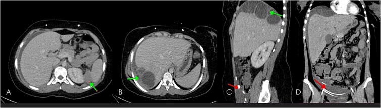
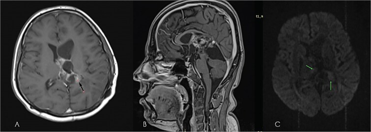
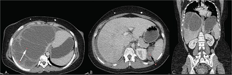
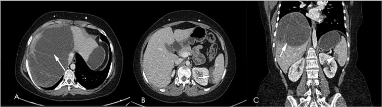
High-grade gliomas (HGGs) are aggressive primary brain tumors with a poor prognosis. Although extracranial metastasis is uncommon, peritoneal dissemination via ventriculoperitoneal (VP) shunts is extremely rare. Here, we describe the case of an 18-year-old female with HGG who developed peritoneal metastasis one year after VP shunt placement. Although VP shunting in the presence of an intracranial high-grade tumor is generally not contraindicated, shunt-related metastasis should be recognized as a potential risk and an important, albeit rare, clinical presentation. This case highlights the diagnostic challenges and therapeutic limitations of this rare complication. Understanding this phenomenon is crucial for developing effective monitoring and treatment strategies.

Keywords: Extraneural metastasis; Ventriculoperitoneal shunt; case report; glioma; peritoneal metastasis.


Conflict of interest statement

No conflicts of interest.

Figures

Figure 1
Figure 1
Pre-operative brain MRI of diffuse astrocytoma: T2WI axial (A), & sagittal (B), T1WI axial (C), FLAIR axial (D), ADC axial (E) images showing bubbly heterogeneously enhancing mass (enhanced images not shown), mixed signal intensity in T2WI, iso in T1WI, high on FLAIR, showing diffusion restriction on ADC; indicating highly cellular high-grade tumor involving deep white matter, extending to pineal and quadrigeminal cisterns causing mass effect with moderate hydrocephalous.
Figure 2
Figure 2
(A) Brain biopsy showed a diffuse cellular lesion with medium power magnification. (B) Brain biopsy showed a cellular lesion featuring marked nuclear atypia and pleomorphism and fibrillary cytoplasm, high power magnification. (C) Brain biopsy, and immunohistochemical staining for GFAP showed a strong and diffuse pattern of stain of the neoplastic cells. (D) Brain biopsy, ki67 immunohistochemical staining showed a high proliferative index.
Figure 3
Figure 3
Early post-operative brain MRI images (A) axial T1WI without contrast, (B & C) axial T2WI; showing small hematoma at operative bed (green arrow in A), & small LT subdural hematoma (green arrow in B), with shunt tube seen at improper site at RT cauda-thalamic recess (black arrow in B), another functioning properly seated shunt tube seen within RT lateral ventricle sagittal T2WI in (D) showing residual cystic component of the mass (diffuse astrocytoma) green arrow in the Quadrigeminal and pineal region cisterns.
Figure 4
Figure 4
Follow-up brain MRI, axial (A), sagittal (B), post-contrast images, sagittal T2WI (C), and ADC map (D) showing non-enhancing cystic lesion with free diffusion representing residual disease and getting smaller in size in response to CCRT as compared with previous images. Note the resolution of early post-operative hematoma and subdural hemorrhage.
Figure 5
Figure 5
Computed tomography post contrast portal venous phase images axial (A, B), sagittal (C) and coronal reformat (D), showing large thick ascites as well as enhancing peritoneal cystic masses (green arrows) representing metastatic peritoneal lesions. Note the Ventriculoperitoneal shunts (in red arrows).
Figure 6
Figure 6
Diagnostic laparoscopy which showed multiple peritoneal and omental masses with huge ascites.
Figure 7
Figure 7
(A) A peritoneal biopsy showed a cellular spindle and low power magnification. (B) A peritoneal biopsy showed a cellular spindle lesion with areas of palisading necrosis and microvascular proliferation, medium power magnification. (C) Peritoneal biopsy showing cellular lesion with marked atypia and pleomorphism and foci of microvascular proliferation, high power magnification. (D) Peritoneal biopsy, and immunohistochemical staining for GFAP showed a strong and diffuse positivity of the neoplastic cells.
Figure 8
Figure 8
Further follow-up (14 months from operation) brain MRI: post-contrast images; axial (A), Sagittal (B), and diffusion-weighted image (C), showing disease progression with enlarging peripherally enhancing cystic lesion with mild diffusion restriction. Note: mass effect causing dilatation of frontal horn of LT lateral ventricle.
Figure 9
Figure 9
Computed tomography post-contrast portal venous phase images axial (A, B), and coronal reformat (C), showing interval progression in size of enhancing peritoneal cystic masses (white arrow in A, black in B) representing metastatic peritoneal lesions.
Figure 10
Figure 10
Computed tomography post-contrast portal venous phase images axial (A, B), and coronal reformat (C), dated august 2024; showing slight regression in size of enhancing peritoneal cystic metastatic lesions (white arrows in A & B, arrowhead in C) response less than 30%, regarded as stable disease per RECIST criteria.

References

    1. Wu J, Al-Zahrani A, Beylerli O. et al. Circulating miRNAs as diagnostic and prognostic biomarkers in high-grade gliomas. Front Oncol 2022;12:898537. 10.3389/fonc.2022.898537 - DOI - PMC - PubMed
    1. Prabhakaran N, Miller DC, Litofsky NS. et al. Extraneural metastasis of primary glioma occurring in a setting of occupational ionizing radiation exposure. Case Rep Neurol Med 2019;2019:1748739. - PMC - PubMed
    1. Pasquier B, Pasquier D, N'golet A. et al. Extraneural metastases of astrocytomas and glioblastomas: Clinicopathological study of two cases and review of literature. Cancer 1980;45:112–25. - PubMed
    1. Stephens S, Tollesson G, Robertson T. et al. Diffuse midline glioma metastasis to the peritoneal cavity via ventriculoperitoneal shunt: case report and review of literature. J Clin Neurosci 2019;67:288–93. - PubMed
    1. National Comprehensive Cancer Network. Central Nervous System Cancers (Version 3.2024). Plymouth Meeting, PA: National Comprehensive Cancer Network; 2024. Available at: https://www.nccn.org/professionals/physician_gls/pdf/cns.pdf. Accessed October 7, 2024.

Publication types

LinkOut - more resources