

Granular Cell Ameloblastomatous Transformation From the Remnants of a Dentigerous Cyst: A Unique Case Report
- PMID: 40585620
- PMCID: PMC12205571
- DOI: 10.7759/cureus.85021
Granular Cell Ameloblastomatous Transformation From the Remnants of a Dentigerous Cyst: A Unique Case Report
Abstract
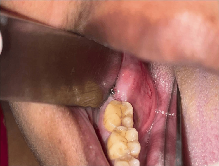
Granular cell ameloblastoma is a unique, infrequent histologic variant of unicystic/multicystic ameloblastoma showing distinct histologic and immunohistochemical features. The prognosis and treatment are similar to other common subtypes of solid or multicystic ameloblastoma. Granular cell ameloblastoma should be distinguished from other lesions with granular cells mainly due to its high risk of recurrence. Although it is rare, it has greater recurrence potential and chances of malignant potential. A better knowledge of the molecular pathogenesis of ameloblastoma and its various subtypes may provide diagnostic and therapeutic benefits. We are reporting a case of granular cell ameloblastoma arising from the wall of a dentigerous cyst. The lining of the dentigerous cyst shows a potential for neoplastic transformation to ameloblastoma, squamous cell carcinoma, and mucoepidermoid carcinoma.
Keywords: dentigerous cyst; enucleation; granular cell ameloblastoma; lysosomal aggregation; neoplasm.
Copyright © 2025, Hebbale et al.
Conflict of interest statement
Human subjects: Consent for treatment and open access publication was obtained or waived by all participants in this study. Conflicts of interest: In compliance with the ICMJE uniform disclosure form, all authors declare the following: Payment/services info: All authors have declared that no financial support was received from any organization for the submitted work. Financial relationships: All authors have declared that they have no financial relationships at present or within the previous three years with any organizations that might have an interest in the submitted work. Other relationships: All authors have declared that there are no other relationships or activities that could appear to have influenced the submitted work.
Figures


References
-
- Odontogenic cysts. Rajendra Santosh AB. Dent Clin North Am. 2020;64:105–119. - PubMed
-
- Odontogenic cysts: classification, histological features and a practical approach to common diagnostic problems. Brown SJ, Conn BI. https://doi.org/10.1016/j.mpdhp.2022.02.007 Diagn Histopathol. 2022;28:253–266.
-
- Odontogenic cysts and tumors. Chi AC, Neville BW. Surg Pathol Clin. 2011;4:1027–1091. - PubMed
-
- Primary intraosseous squamous cell carcinoma arising in odontogenic cysts: a report of five cases and a review of the literature. Wolk DR, Freedman DP, Reich DR. Oral Surg Oral Med Oral Pathol Oral Radiol. 2022;133:0.
Publication types
LinkOut - more resources
Full Text Sources