. 2025 Jun 2:38:13971.
doi: 10.3389/ti.2025.13971. eCollection 2025.

Delivery of a Muscle-Targeted Adeno-Associated Vector Via Ex Vivo Normothermic Perfusion Is Efficient, Durable, and Safe in a Preclinical Porcine Heart Transplant Model

Affiliations

Delivery of a Muscle-Targeted Adeno-Associated Vector Via Ex Vivo Normothermic Perfusion Is Efficient, Durable, and Safe in a Preclinical Porcine Heart Transplant Model

Krish C Dewan et al. Transpl Int. .

Abstract

Normothermic ex-vivo organ perfusion (EVP) systems not only provide a physiological environment that preserves donor organ function outside the body but may also serve as platforms for ex-vivo organ modification via gene therapy. In this study, we demonstrated that a rationally designed muscle-tropic recombinant AAV, AAV-SLB101, delivered to the donor heart during brief normothermic EVP achieves durable cardiac transgene expression out to 90 and 120 days post-transplant in a porcine preclinical model. Moreover, transgene expression was detectable as early as 48 h post-transplant. Histological and MRI analyses of the donor myocardium showed no functional or structural impact on the allograft and no off-target gene expression in the recipient. This work will serve as a critical foundation to inform translational studies with therapeutic transgenes to improve allo-, xeno-, and auto-heart transplant outcomes.

Keywords: adeno-associated virus vector; ex vivo heart preservation; gene therapy; heart transplantation; transgene durability.


Conflict of interest statement

CM has received stock compensation for serving as a consult for TransMedics Inc. WC, MB, KP, and AC are employees and shareholders of Solid Biosciences Inc. PL is an employee of TransMedics, Inc. The remaining authors declare that the research was conducted in the absence of any commercial or financial relationships that could be construed as a potential conflict of interest.

Figures

FIGURE 1
FIGURE 1
Overview study design of animal experiments and analysis pipeline.
FIGURE 2
FIGURE 2
Vector DNA over time. Viral genome copy numbers per µg DNA in each chamber of the transplanted and native hearts at various timepoints after transplantation (A) 1 day (B) 2 days (C) 90 days, and (D) 120 days. Samples are labeled along with the number of the cross section from which they were obtained. Cross sections are numbered 1–5 from base/atrium of the heart to apex respectively. All samples were obtained from the medial aspect of each chamber for consistency. (E) Average vector copy number across the entire allograft at 24 h, 48 h, 90 days and 120 days. (F) Negligible viral DNA at any timepoint in any of the native untreated hearts. (G) Side-by-side comparison of vector DNA copy numbers by chamber over time. LA, left atrium; SA, atrial septum; RA, right atrium; LV, left ventricle; RV, right ventricle; IVS, interventricular septum; NTC, no template control.
FIGURE 3
FIGURE 3
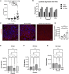
Luciferase expression throughout the myocardium. Luciferase expression at (A) 1 day (B) 2 days (C) 90 days and (D) 120 days expressed as relative light units (RLU) per ug tissue protein. Transgene expression is displayed both on the graphs numerically as well as in the heatmap. Graphs are plotted on the log10 scale. (E) Representative histology with immunofluorescent staining of luciferase(red) and counter staining the nucleus with DAPI (blue). All images are of medial wall from the third cross-section at ×20 magnification, with the scale bar indicating 100 μm. LA, left atrium; SA, atrial septum; RA, right atrium; LV, left ventricle; RV, right ventricle; IVS, interventricular septum. Nomenclature of chamber: chamber, cross section, wall (e.g., LV2a was a left ventricular sample from the anterior aspect of cross section 2).
FIGURE 4
FIGURE 4
Luciferase biodistribution and progression of expression over time. (A) Overall luciferase expression from 1 to 120 days assessed by luciferase activity assay. (B) Luciferase expression over time by chamber. Graphs are plotted on the log10 scale. (C) Representative luciferase staining of endomyocardial biopsies. (D) Mean fluorescent intensity (MFI) per nucleus of luciferase among endomyocardial biopsy samples at 30, 60, and 90 days. Luciferase enzymatic activity across all chambers (atria and ventricles) at all three timepoints from 2 days (E) to 90 days (F) to 120 days (G) post-transplant. RLU values were calculated by pooling values for all samples from each chamber (n = 3 atria, n = 10 LV, n = 9 IVS, n = 9, IVS). LV, left ventricle; IVS, interventricular septum; RV, right ventricle.
FIGURE 5
FIGURE 5
Luciferase expression in extra-cardiac organs. (A) Luciferase immunofluorescent staining of recipient extra-cardiac organs including the liver, lung, or skeletal muscle at any timepoint after transplantation. All images are at ×20 magnification, with the scale bar indicating 100 μm. Psoas m. = psoas muscle (representative of skeletal muscle). (B) Viral copy numbers per ug DNA in the liver and psoas muscle at 24 h, 48 h, 90 days, and 120 days (C) Luciferase activity at all timepoints in the lung, liver, and skeletal muscle.
FIGURE 6
FIGURE 6
Safety of ex vivo vector delivery via cardiac MRI and histopathologic assessment. (A) Representative preoperative and postoperative (day 12) CMR T1 mapping and DE-CMR images. (B) Change in left ventricular mass in the native heart (C) Change in left ventricular mass in the allograft (C). (D) Changes in T1 enhancement in the recipient native heart (E) Changes in T1 enhancement in the allograft. (F) Extracellular volume (ECV) in the allograft. H&E staining of the recipient’s allograft (G) native heart (H), liver (I), and lung tissue (J).

References

    1. Khush KK, Cherikh WS, Chambers DC, Harhay MO, Hayes D, Jr, Hsich E, et al. The International Thoracic Organ Transplant Registry of the International Society for Heart and Lung Transplantation: Thirty-Sixth Adult Heart Transplantation Report - 2019; Focus Theme: Donor and Recipient Size Match. J Heart Lung Transpl (2019) 38(10):1056–66. 10.1016/j.healun.2019.08.004 - DOI - PMC - PubMed
    1. Bryner BS, Schroder JN, Milano CA. Heart Transplant Advances: Ex vivo Organ-Preservation Systems. JTCVS Open (2021) 8:123–7. 10.1016/j.xjon.2021.04.020 - DOI - PMC - PubMed
    1. Qin G, Jernryd V, Sjöberg T, Steen S, Nilsson J. Machine Perfusion for Human Heart Preservation: A Systematic Review. Transpl Int (2022) 35:10258. 10.3389/ti.2022.10258 - DOI - PMC - PubMed
    1. Mendiola Pla M, Chiang Y, Roan J-N, Bowles DE. Gene Therapy for Cardiac Transplantation. London, United Kingdom: IntechOpen; (2022).
    1. Mendiola PM, Evans A, Lezberg P, Bowles DE. Ex vivo Delivery of Viral Vectors by Organ Perfusion for Cardiac Transplantation Gene Therapy. Methods Mol Biol (2022) 2573:249–59. 10.1007/978-1-0716-2707-5_19 - DOI - PubMed

LinkOut - more resources