. 2024 Jun;4(6):2300101.
doi: 10.1002/anbr.202300101. Epub 2024 Apr 26.

A Microfluidic Strategy to Capture Antigen-Specific High-Affinity B Cells

Affiliations

A Microfluidic Strategy to Capture Antigen-Specific High-Affinity B Cells

Ahmed M Alhassan et al. Adv Nanobiomed Res. 2024 Jun.

Abstract

Assessing B cell affinity to pathogen-specific antigens prior to or following exposure could facilitate the assessment of immune status. Current standard tools to assess antigen-specific B cell responses focus on equilibrium binding of the secreted antibody in serum. These methods are costly, time-consuming, and assess antibody affinity under zero force. Recent findings indicate that force may influence BCR-antigen binding interactions and thus immune status. Herein, a simple laminar flow microfluidic chamber in which the antigen (hemagglutinin of influenza A) is bound to the chamber surface to assess antigen-specific BCR binding affinity of five hemagglutinin-specific hybridomas from 65 to 650 pN force range is designed. The results demonstrate that both increasing shear force and bound lifetime can be used to enrich antigen-specific high-affinity B cells. The affinity of the membrane-bound BCR in the flow chamber correlates well with the affinity of the matched antibodies measured in solution. These findings demonstrate that a microfluidic strategy can rapidly assess BCR-antigen-binding properties and identify antigen-specific high-affinity B cells. This strategy has the potential to both assess functional immune status from peripheral B cells and be a cost-effective way of identifying individual B cells as antibody sources for a range of clinical applications.

Keywords: affinities; avidities; cell separations; immunology.


Conflict of interest statement

The authors declare no conflict of interest.

Figures
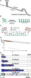
Figure 1
Figure 1
A) Schematic of antigen functionalized on the surface of the device and B) cells binding to the antigen‐coated surface. C) COMSOL Multiphysics model of cells entering the microfluidic device and flowing horizontally and D) settling to the bottom along the length of the device at different flow rates. Cells that contact the surface (blue) are frozen in place.
Figure 2
Figure 2
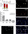
A) Schematic demonstrating how the SwHEL mouse is created with ≈20% of the cells from the spleen at HEL‐specific B cells (produced in part from BioRender). B) Flow cytometry showing subset of spleen cells that are specific to HEL from WT and SwHEL mice. C) Capture efficiency at 100 μL h−1 (n = 3, mean ± SEM) of WT and SwHEL cells in microfluidic device coated with different concentrations of HEL and D) microscopic imaging showing cells under shear stress of 0.30 dyn cm−2 in the device. Circled cells are bound to HEL.
Figure 3
Figure 3
A) Schematic demonstrating how hybridoma technology produces monoclonal hybridoma clones (produced in part from BioRender). B) Optical Density binding curve generated by ELISA assay fitted to measure antibody affinity. Groups with different letters are significantly different (p < 0.05). C) Antibody affinity to HA (ND: not detected), and D) avidity to influenza virion measured by OI‐RD.
Figure 4
Figure 4
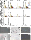
A) Capture efficiency (n ≥ 3, ± SEM) of five HA‐specific hybridoma lines and negative control under shear stress of 0.03, 0.06, and 0.15 dyn cm−2 with minimum binding criteria of 10, 20, 50, and 100 s. Groups with different letters are significantly different (p < 0.05). B) Microscopic imaging showing cells under shear stress of 0.06 dyn cm−2 in the device. Circled cells are bound to HA.
Figure 5
Figure 5
A) Composition of hybridoma cell lines in the mixture that is perfused in the device. B) Fold change in enrichment (Bound Cells (%)/Perfusate (%)) for different cell populations in mixture when perfused at 10 and 20 μL h−1 (n = 3, mean ± SEM). *significantly different from other groups (p < 0.05). C–G) Microscopic imaging showing cells under shear stress of 0.06 dyn cm−2. H36 (yellow), H163 (red), H37 (blue), and DS.1, H35 and H143 (cyan) are shown individually and in the mixture in which they were perfused. Circled cells are bound to HA. Bound cells were measured according to a minimum binding criterion of 10 s. Values that differ from 1‐fold change with statistical significance (p < 0.05) are indicated.
Figure 6
Figure 6
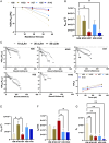
A) Data fit based on first‐order kinetic model to measure effective k on. B) Measured values of effective k on for the different hybridoma cell lines. C) Survival curve fits to determine force‐dependent k off, where bound lifetime is the length of time the cells remain bound for more than 10 s and D) data fit to determine reactive compliance (x β ) and effective k off at zero‐force. E) Values of effective k off at zero‐force and F) reactive compliance (x β ). G) Membrane‐bound BCR affinity (effective konkoff) to HA. *significantly different (p < 0.05); **significantly different (p < 0.01).

Update of

References

    1. Victora G. D., Nussenzweig M. C., Annu. Rev. Immunol. 2022, 40, 413. - PubMed
    1. Wong R., Belk J. A., Govero J., Uhrlaub J. L., Reinartz D., Zhao H., Errico J. M., D’Souza L., Ripperger T. J., Nikolich‐Zugich J., Shlomchik M. J., Satpathy A. T., Fremont D. H., Diamond M. S., Bhattacharya D., Immunity 2020, 53, 1078. - PMC - PubMed
    1. Weisel F. J., Zuccarino‐Catania G. V., Chikina M., Shlomchik M. J., Immunity 2016, 44, 116. - PMC - PubMed
    1. LeBien T. W., Tedder T. F., Blood J. Am. Soc. Hematol. 2008, 112, 1570. - PMC - PubMed
    1. Doucett V. P., Gerhard W., Owler K., Curry D., Brown L., Baumgarth N., J. Immunol. Methods 2005, 303, 40. - PubMed

LinkOut - more resources