. 2025 Jun 26;17(6):106488.
doi: 10.4252/wjsc.v17.i6.106488.

Mesenchymal stem cells-derived exosomes alleviate radiation induced pulmonary fibrosis by inhibiting the protein kinase B/nuclear factor kappa B pathway

Affiliations

Mesenchymal stem cells-derived exosomes alleviate radiation induced pulmonary fibrosis by inhibiting the protein kinase B/nuclear factor kappa B pathway

Li-Li Wang et al. World J Stem Cells. .

Abstract

Background: Radiation induced pulmonary fibrosis (RIPF) is a long-term lung condition with a bleak outlook and few treatment possibilities. Mesenchymal stem cells (MSCs)-derived exosomes (MSCs-exosomes) possess tissue repair and regenerative properties, but their exact mechanisms in RIPF remain unclear. This study explores whether MSCs-exosomes can alleviate RIPF by modulating inflammation, extracellular matrix (ECM) accumulation, and epithelial-mesenchymal transition (EMT) via the protein kinase B (Akt)/nuclear factor kappa B (NF-κB) pathway.

Aim: To assess the therapeutic potential and mechanisms of MSCs-exosomes in RIPF.

Methods: Sprague-Dawley rats were received 30 Gy X-ray radiation on the right chest to induce RIPF, while RLE-6TN and BEAS-2B cell lines were exposed to 10 Gy X-rays. Using differential centrifugation, MSCs-exosomes were isolated, and their protective effects were examined both in vivo and in vitro. Inflammatory cytokine concentrations were measured using Luminex liquid chip detection and enzyme linked immunosorbent assay. ECM and EMT-related proteins were analyzed using immunohistochemistry, western blotting, and real-time quantitative polymerase chain reaction. Western blotting and immunohistochemistry were also used to investigate the mechanisms underlying MSCs-exosomes' effects in RIPF.

Results: Administration of MSCs-exosomes significantly mitigated RIPF, reduced collagen deposition, and decreased levels of various inflammatory cytokines. Additionally, MSCs-exosomes prevented radiation-induced ECM accumulation and EMT. Treatment with MSCs-exosomes notably promoted cell proliferation, suppressed inflammation, and reversed ECM deposition and EMT in radiation-exposed alveolar epithelial cells. Mechanistic analysis further revealed that MSCs-exosomes exerted their anti-RIPF effects by inhibiting the Akt/NF-κB pathway, as shown in both in vivo and in vitro models.

Conclusion: MSCs-exosomes mitigate RIPF by suppressing inflammation, ECM deposition, and EMT through Akt/NF-κB inhibition, highlighting their potential as a therapeutic strategy.

Keywords: Exosomes; Mesenchymal stem cells; Nuclear factor kappa B; Protein kinase B; Radiation induced pulmonary fibrosis.


Conflict of interest statement

Conflict-of-interest statement: All the authors report no relevant conflicts of interest for this article.

Figures
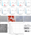
Figure 1
Figure 1
Identification of mesenchymal stem cells and mesenchymal stem cells-derived exosomes. A: Identification of mesenchymal stem cells surface marker proteins (CD105, CD90, CD73, D45, CD34, CD14, CD19 and HLA-DR) by flow cytometry; B: The morphology (× 100) of mesenchymal stem cells, and identification of osteogenic (× 100), chondrogenic (× 100) and adipogenic (× 100) differentiation abilities of mesenchymal stem cells; C: Electron microscopic image of exosomes; D: Particle diameter of exosomes detected by nanoparticle tracking analysis; E: Western blot assay utilized for the detection of the surface marker proteins (ALIX, tumor susceptibility gene 101 and CD9) of exosomes (n = 1). EXO: Exosomes; MSCs: Mesenchymal stem cells; TSG101: Tumor susceptibility gene 101.
Figure 2
Figure 2
Therapeutic efficacy of mesenchymal stem cells-derived exosomes in radiation induced pulmonary fibrosis rats. A: Schematic diagram of the strategy for radiation induced pulmonary fibrosis in rats; B: Changes of body weight of rats in each group (n = 6); C: Computed tomography images of rat lungs and morphological changes in lung tissue; D: Hematoxylin and eosin staining and Masson staining of lung tissue among different groups; E: Quantification of fibrosis by Ashcroft score (n = 6); F: Quantification of area percentage of collagen (n = 6); G: Hydroxyproline content in lung tissues (n = 6). aP < 0.05 vs control + phosphate buffered saline, bP < 0.01 vs control + phosphate buffered saline, cP < 0.0001 vs control + phosphate buffered saline, dP < 0.0001 vs irradiation + phosphate buffered saline. CON: Control; PBS: Phosphate buffered saline; IR: Irradiation; EXO: Exosomes; HYP: Hydroxyproline.
Figure 3
Figure 3
Mesenchymal stem cells-derived exosomes ameliorated radiation induced inflammation in radiation induced pulmonary fibrosis rats. A-F: Changes in the level of inflammatory cytokines [interleukin (IL)-1β, IL-2, IL-6, IL-12p70, interferon-γ and tumor necrosis factor-α] of serum samples collected at different time points in three groups (n = 6); G: The total protein concentration in bronchoalveolar lavage fluid of rats at 16 weeks (n = 6); H and I: Changes in inflammatory factors (IL-6 and tumor necrosis factor-α) in rat bronchoalveolar lavage fluid at 16 weeks (n = 6); J: Real-time quantitative polymerase chain reaction detection of transforming growth factor-β1, IL-6 and IL-1β mRNA levels in rat lung tissue (n = 6); K and L: Immunohistochemical staining to observe the staining intensity of transforming growth factor-β1, IL-6, IL-1β, CD3 and CD68 in rat lung tissue. aP < 0.05 vs control + phosphate buffered saline, bP < 0.01 vs control + phosphate buffered saline, cP < 0.001 vs control + phosphate buffered saline, dP < 0.0001 vs control + phosphate buffered saline, eP < 0.05 vs irradiation + phosphate buffered saline, fP < 0.01 vs irradiation + phosphate buffered saline, gP < 0.001 vs irradiation + phosphate buffered saline, hP < 0.0001 vs irradiation + phosphate buffered saline. CON: Control; IR: Irradiation; EXO: Exosomes; PBS: Phosphate buffered saline; IL: Interleukin; IFN: Interferon; TNF: Tumor necrosis factor; TGF: Transforming growth factor; BALF: Bronchoalveolar lavage fluid.
Figure 4
Figure 4
Mesenchymal stem cells-derived exosomes suppressed radiation induced extracellular matrix and epithelial-mesenchymal transition in radiation induced pulmonary fibrosis rats. A: Immunohistochemical staining to observe the staining intensity of E-cadherin, vimentin, alpha-smooth muscle actin (α-SMA) and collagen type 1 alpha 1 (COL1A1) in rat lung tissue; B and C: Western blotting detection of E-cadherin, vimentin, α-SMA and COL1A1 protein expression levels in rat lung tissue (n = 3); D: Real-time quantitative polymerase chain reaction detection of E-cadherin, vimentin, α-SMA and COL1A1 mRNA levels in rat lung tissue (n = 6). aP < 0.05 vs control + phosphate buffered saline, bP < 0.01 vs control + phosphate buffered saline, cP < 0.0001 vs control + phosphate buffered saline, dP < 0.05 vs irradiation + phosphate buffered saline, eP < 0.001 vs irradiation + phosphate buffered saline, fP < 0.0001 vs irradiation + phosphate buffered saline. CON: Control; PBS: Phosphate buffered saline; IR: Irradiation; EXO: Exosomes; α-SMA: Alpha-smooth muscle actin; COL1A1: Collagen type 1 alpha 1.
Figure 5
Figure 5
Mesenchymal stem cells-derived exosomes promoted proliferation and suppressed inflammation, extracellular matrix, epithelial-mesenchymal transition in radiation induced alveolar epithelial cells. A and B: The cell proliferations of RLE-6TN and BEAS-2B cells were detected by CCK8 assay. OD value (450 nm) was measured at 24 hours, 48 hours and 72 hours (n = 3); C and D: The secretion levels of inflammatory chemokines [tumor necrosis factor-α, interleukin (IL)-6, IL-1β, IL-10] in the culture supernatants were measured by enzyme linked immunosorbent assay (n = 3); E and F: The mRNA expressions of E-cadherin, vimentin, alpha-smooth muscle actin and collagen type 1 alpha 1 were detected by real-time quantitative polymerase chain reaction in RLE-6TN and BEAS-2B cells (n = 3). aP < 0.05 vs control + phosphate buffered saline, bP < 0.01 vs control + phosphate buffered saline, cP < 0.001 vs control + phosphate buffered saline, dP < 0.0001 vs control + phosphate buffered saline, eP < 0.05 vs irradiation + phosphate buffered saline, fP < 0.01 vs irradiation + phosphate buffered saline, gP < 0.001 vs irradiation + phosphate buffered saline, hP < 0.0001 vs irradiation + phosphate buffered saline. CON: Control; PBS: Phosphate buffered saline; IR: Irradiation; EXO: Exosomes; IL: Interleukin; TNF: Tumor necrosis factor; α-SMA: Alpha-smooth muscle actin; COL1A1: Collagen type 1 alpha 1.
Figure 6
Figure 6
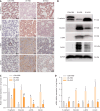
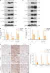
Mesenchymal stem cells-derived exosomes inhibited protein kinase B/nuclear factor kappa B signaling in radiation induced pulmonary fibrosis. A-D: Western blot analysis of protein kinase B (Akt)/nuclear factor kappa B (NF-κB) signaling pathway expression levels in rat lung tissue at 16 weeks (n = 3) (A and B); western blot analysis of Akt/NF-κB signaling pathway expression levels in RLE-6TN cells (n = 3) (C and D); E and F: Immunohistochemistry was used to determine the staining intensity of p-Akt, p-IkappaB kinase α/β and p-NF-κB p65 in rat lung tissue at 16 weeks (n = 6). aP < 0.0001 vs control + phosphate buffered saline, bP < 0.01 vs control + phosphate buffered saline, cP < 0.0001 vs irradiation + phosphate buffered saline. CON: Control; IR: Irradiation; PBS: Phosphate buffered saline; EXO: Exosomes; Akt: Protein kinase B; IKK: IkappaB kinase; NF-κB: Kinase nuclear factor kappa B.

Similar articles

References

    1. Zhang Z, Zhou J, Verma V, Liu X, Wu M, Yu J, Chen D. Crossed Pathways for Radiation-Induced and Immunotherapy-Related Lung Injury. Front Immunol. 2021;12:774807. - PMC - PubMed
    1. Hanania AN, Mainwaring W, Ghebre YT, Hanania NA, Ludwig M. Radiation-Induced Lung Injury: Assessment and Management. Chest. 2019;156:150–162. - PMC - PubMed
    1. Li J, Wang R, Shi W, Chen X, Yi J, Yang X, Jin S. Epigenetic regulation in radiation-induced pulmonary fibrosis. Int J Radiat Biol. 2023;99:384–395. - PubMed
    1. Chen Z, Wu Z, Ning W. Advances in Molecular Mechanisms and Treatment of Radiation-Induced Pulmonary Fibrosis. Transl Oncol. 2019;12:162–169. - PMC - PubMed
    1. Huang C, Ogawa R. Role of Inflammasomes in Keloids and Hypertrophic Scars-Lessons Learned from Chronic Diabetic Wounds and Skin Fibrosis. Int J Mol Sci. 2022;23:6820. - PMC - PubMed

LinkOut - more resources