. 2025 Jun 9;15(14):6938-6956.
doi: 10.7150/thno.107659. eCollection 2025.

Enhancing hair regrowth using rapamycin-primed mesenchymal stem cell-derived exosomes

Affiliations

Enhancing hair regrowth using rapamycin-primed mesenchymal stem cell-derived exosomes

Manju Shrestha et al. Theranostics. .

Abstract

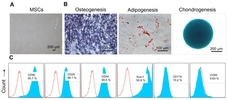
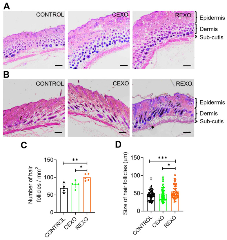
Rationale: Hair loss affects millions globally, with limited effective treatments available and significant psychological impacts. Mesenchymal stem cells (MSCs) and MSC-derived exosomes hold therapeutic potential by modulating cellular communication, reducing inflammation, and supporting hair follicular regeneration. Rapamycin, a mechanistic target of rapamycin (mTOR) inhibitor, enhances MSC therapeutic potential by promoting the release of growth factors and signaling molecules. Thus, this study explores the benefit of priming effect of rapamycin on enhancing the function of MSC-derived exosomes to promote hair regrowth in a depilation-induced murine model. Methods: MSCs were primed with rapamycin, and exosomes were extracted from the MSC-conditioned media using ultrafiltration and poly (ethylene glycol) (PEG) precipitation. Dermal fibroblasts were treated with several doses of exosomes to evaluate the in vitro effect of rapamycin-primed MSC-derived exosomes (REXO). The depilated mice were administered exosomes via intradermal route and the hair regrowth was monitored for 15 days, followed by gene expression analysis and histological examination. Results: Dermal fibroblasts treated with REXO showed a higher proliferation rate and an increase in genes related to Wnt/β-catenin signaling, autophagy, and growth factors compared to non-primed MSC-derived exosomes (CEXO). In vivo REXO therapy via intradermal injection to the depilated areas in mice enhanced hair follicle development, hair density, and hair activation markers compared with the control and naive exosome treatments. Conclusion: REXO therapy effectively enhances hair regrowth thus this approach could offer a clinically effective therapy for hair loss treatment.

Keywords: Wnt/β-catenin signaling; exosome; hair regrowth; mesenchymal stem cell; rapamycin.


Conflict of interest statement

Competing Interests: The authors have declared that no competing interest exists.

Figures

Figure 1
Figure 1
Engineering of MSCs with rapamycin (R) to enhance exosome (EXO) release and function, promoting hair regrowth in an experimental mouse model. REXO therapy exhibited its therapeutic effects via modulation of Wnt/β-catenin signaling and autophagy, thereby promoting hair follicle activation and effective hair regrowth. (The figure was designed with Biorender.com).
Figure 2
Figure 2
Characterization of MSCs isolated from mouse adipose tissues. (A) Representative bright-field image showing the morphology of MSCs isolated from mouse adipose tissue. (B) Representative images showing MSCs differentiated into osteocytes, adipocytes, and chondrocytes, as evidenced by Alkaline Phosphatase, Oil Red O, and Alcian blue staining, respectively. (C) Surface marker profiling of MSCs by flow cytometry. MSCs showed higher levels of CD90, CD29, CD44, and Sca-1, and lower levels of CD11b and CD45. The blue and red curves indicate samples stained with target antibodies and isotype control antibodies, respectively.
Figure 3
Figure 3
Isolation and characterization of exosomes derived from control MSCs (CEXO) and exosomes derived from rapamycin-primed MSCs (REXO). (A) Schematic illustration of the procedure followed for isolating the exosomes, created by biorender.com (B) Size distribution of EXO as measured by NTA. (C) Morphology of exosomes according to TEM imaging (scale bar, 100 nm). (D) Assessment of exosomal surface markers based on western blot analysis. (E) Purity of exosomes determined by NTA. Data are expressed as mean ± SD (n = 5) and were analyzed using unpaired two-tailed t-test. *p < 0.05, ** p < 0.01.
Figure 4
Figure 4
In vitro effect of exosomes on dermal fibroblasts. (A) Illustration for treatment of exosomes for different analysis. Assessment of dermal cell viability after treatment with exosomes as determined by (B) live/dead staining assay, scale bars = 200 µm and (C) CCK-8 assay. (D) Assessment of dermal cell proliferation in response to exosome treatment, with results expressed as cell numbers compared to control. (E) Gene expression levels as evaluated by qRT-PCR, showing the gene expression levels linked to Wnt/β-catenin signaling (Wnt10b, Wnt5a, Wnt1a, and β-catenin), autophagy (Beclin-1, LC3A, and LC3B), and growth factors (VEGF-A and PDGF-B) in dermal fibroblasts treated with exosomes. (F) Representative images of the DiI-labeled CEXO and REXO into dermal fibroblasts (Red: DiI labeled EXOs, Green: Celltracker Green CMFDA). Scale bar = 100 µm (G) Quantification of exosomes uptake by dermal fibroblasts. Data are expressed as mean ± SD (n = 3) and were analyzed using an unpaired two-tailed t-test for comparing two groups, and one-way ANOVA with Tukey's multiple comparisons test for comparing more than 3 groups. * p < 0.05, ** p < 0.01, *** p < 0.001, ****p < 0.0001, and ns = not significant.
Figure 5
Figure 5
Enhanced hair growth induced by rapamycin-primed MSC-derived exosomes (REXO) in an in vivo mouse model. (A) Schematic of the hair growth model timeline, showing the key time points for exosome administration and evaluation. (B) Images of mice on days 0, 3, 7, 10, 11,12, 13, 14 and 15 post-exosome treatment, illustrating hair growth progression (n = 6). (C) Percentage of hair growth in mice (as determined by ImageJ). Data are represented as mean ± SD (n = 6 per group) and were analyzed by two-way ANOVA followed by Tukey's multiple comparisons test. * p < 0.05, ** p < 0.01, *** p < 0.001 and **** p < 0.0001.
Figure 6
Figure 6
REXO enhances hair regrowth by promoting Wnt signaling, autophagy, and growth factors expression. (A) Schematic overview of in vivo sample processing and analysis. (B) Relative mRNA expression levels of key genes associated with Wnt/β-catenin signaling (Wnt-10b, Wnt-5a, Wnt-1a, and β-catenin), autophagy (Beclin-1, LC3A, and LC3B), and growth factors (VEGF-A and PDGF-B) in skin samples extracted from PBS-treated CONTROL, CEXO, and REXO treated mice. (C) Relative protein expressions of the same targets analyzed by western blot. Data are expressed as mean ± SD (n = 6 per group) and were analyzed using one-way ANOVA with Tukey's multiple comparisons test. * p < 0.05, ** p < 0.01, ***p < 0.001, and ****p < 0.0001.
Figure 7
Figure 7
REXO promotes the anagen phase in the hair growth cycle. Representative hematoxylin and eosin (H & E) stained images of skin sections from the mice treated with exosomes showing (A) transverse and (B) longitudinal views. Scale bar = 200 µm. (C) Quantification of number of hair follicles per area. (D) Size distribution of hair follicles. Data are expressed as mean ± SD (n = 5 per group) and were analyzed using one-way ANOVA with Tukey's multiple comparisons test. * p < 0.05, ** p < 0.01 and ***p < 0.001.

References

    1. Phillips TG, Slomiany WP, Allison R. Hair loss: common causes and treatment. Am Fam Physician. 2017;96:371–8. - PubMed
    1. Al Najjar OA, Alkhars MA, Al Molhim SF, AlAjmi MS, Alhafith AA, Al Najjar MA. et al. The impact of androgenic alopecia on the quality of life of male individuals: a cross-sectional study. Cureus. 2023;15:e47760. - PMC - PubMed
    1. Mahfouz MS, Alqassim AY, Hakami FA, Alhazmi AK, Ashiri AM, Hakami AM. et al. Common skin diseases and their psychosocial impact among Jazan population, Saudi Arabia: a cross-sectional survey during 2023. Medicina (Kaunas) 2023;59:1753. - PMC - PubMed
    1. Sattur SS, Sattur IS. Pharmacological management of pattern hair loss. Indian J Plast Surg. 2021;54:422–34. - PMC - PubMed
    1. Asgari AZ, Rufaut NW, Morrison WA, Dilley RJ, Knudsen R, Jones LN. et al. Hair transplantation in mice: challenges and solutions. Wound Repair Regen. 2016;24:679–85. - PubMed

LinkOut - more resources