. 2025 Jun 12;15(14):7025-7044.
doi: 10.7150/thno.117118. eCollection 2025.

A subset of evolutionarily conserved centriolar satellite core components is crucial for sperm flagellum biogenesis

Affiliations

A subset of evolutionarily conserved centriolar satellite core components is crucial for sperm flagellum biogenesis

Bingbing Wu et al. Theranostics. .

Abstract

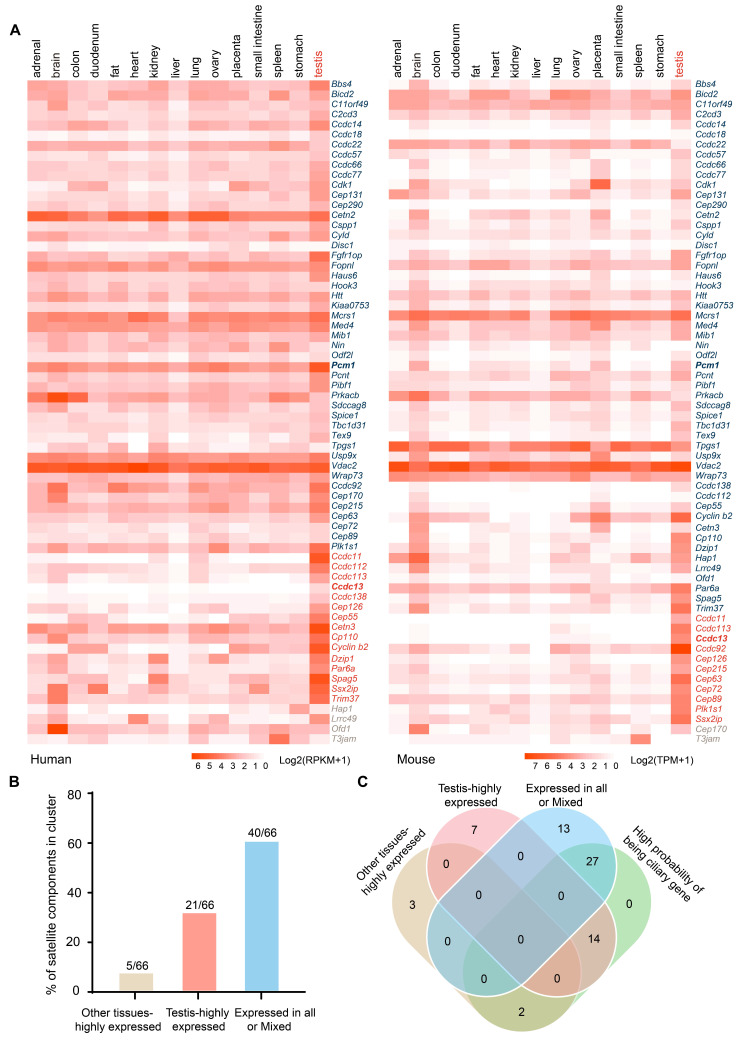
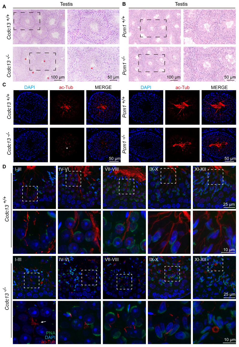
Rationale: Centriolar satellites are non-membranous cytoplasmic granules that cluster around centrosomes, with pericentriolar material 1 (PCM1) serving as the molecular marker for these structures. Although significant progress has been made in understanding their composition, cellular, and organismal functions over the past decades, the tissue-specific roles of centriolar satellite proteins in sperm flagellum biogenesis and male fertility are still not well understood. Methods: We utilize publicly available data and conduct phylogenetic analysis to explore the tissue distribution and conservation of centriole satellite components across flagellated species. Knockout mouse models for Ccdc13 and Pcm1 were constructed to investigate their physiological roles. Sperm morphology and functionality were analyzed using immunofluorescence, transmission electron microscopy, and sperm motility analysis. Immunofluorescence, immunoblotting, co-immunoprecipitation, and proteomics analyses were carried out to elucidate the molecular mechanisms by which CCDC13 regulates sperm flagellum biogenesis. Results: We show that most satellite components are expressed in the testis and associated with ciliary function. Comparative analysis of ciliary-related satellite components across 11 flagellated and non-flagellated species revealed six highly conserved satellite proteins in flagellated species. PCM1, a well-known centriolar satellite scaffolding protein, was found to be less conserved. Based on these findings, we selected CCDC13, a highly conserved satellite protein, and PCM1, a less conserved component, for functional comparison in sperm flagellum biogenesis. Using knockout mouse models, we demonstrated that Ccdc13 deficiency led to male infertility with multiple morphological abnormalities of the sperm flagella (MMAF)-like phenotype due to defects in sperm flagellum biogenesis. While Pcm1 knockout only resulted in decreased sperm motility without affecting flagellum biogenesis. Molecularly, CCDC13 interacts with IMT, IFT-associated proteins, and flagellar components to regulate transport of cargo to proper positions for flagellum biogenesis. Conclusion: This study identifies a subset of highly conserved centriolar satellite proteins essential for sperm flagellum biogenesis. The identification of these proteins provides valuable insights into the genetic mechanisms underlying flagellum function and their evolutionary development. Additionally, defects in these proteins may be associated with male infertility in humans.

Keywords: CCDC13; PCM1; centriolar satellites; flagellum biogenesis; male infertility.


Conflict of interest statement

Competing Interests: The authors have declared that no competing interest exists.

Figures

Figure 1
Figure 1
Expression of most satellite components in testis. (A) Expression heatmaps for 66 centriolar satellite components in human and mouse tissues were generated using publicly available data: the Human Protein Atlas RNA-seq dataset for human tissues and Mouse ENCODE transcriptome data from NCBI. Most centriolar satellite components were detectable in the testis. TissueEnrich was used to categorize genes into three classes: the “Expressed in all or Mixed”, highlighted in blue; the “Testis-highly expressed”, highlighted in red; and the “Other tissues-highly expressed”, highlighted in brown. (B) The percentage of satellite components in different categories based on TissueEnrich analysis. (C) The Venn diagram compares the cilium-related satellite components predicted by the Ciliogenics database with the three classes of satellite components based on their expression characteristics.
Figure 2
Figure 2
A set of highly conserved centriolar satellite proteins might be required for sperm flagellum biogenesis. (A-C) Gene ontology analysis, along with protein-protein interaction networks, was conducted across Homo sapiens, Mus musculus, and Drosophila melanogaster using the STRING database. Satellite proteins involved in ciliogenesis (GO:0060271) are marked by red circles. The thickness of the lines represents the level of support provided by the data. (D) The presence or absence of centrioles and flagella is depicted for 11 organisms representing major eukaryotic lineages in different colors. (Holozoa: violet, Fungi: orange, Plantae: green, Protozoa: blue). Branch lengths are meaningless. Organisms with centrioles and flagella include Homo sapiens, Mus musculus, Gallus gallus, Xenopus laevis, Danio rerio, Drosophila melanogaster, Chlamydomonas reinhardtii, Tetrahymena thermophila, Trypanosoma cruzi. Organisms without centrioles and flagella include Saccharomyces cerevisiae and Arabidopsis thaliana. Protein homologues of satellite components were identified using a BLASTP search in ENSEMBL, NCBI, and UniProt. In all instances, the query sequences were human homologs of these proteins. Based on their distribution patterns across 11 organisms, satellite proteins were classified into four groups and indicated in the right column: Group I—satellite protein orthologues highly conserved in flagellated organisms but absent in non-flagellated ones; Group II—satellite protein orthologues present to varying extents in some flagellated organisms but absent in non-flagellated ones; Group III—satellite protein orthologues less conserved across flagellated species; and Group IV—satellite protein orthologues present in both flagellated and non-flagellated species.
Figure 3
Figure 3
Ccdc13 knockout leads to male infertility, while Pcm1 knockout leads to male subfertility. (A) The different germ-cell stages of spermatogenesis in mice after birth are described. Sg: spermatogonia, Spc: spermatocyte, M, meiotic, RS: round spermatid, ES: elongating spermatid. (B) CCDC13 was expressed starting in P21 testes. TUBULIN served as the loading control. (C) PCM1 was expressed starting in P7 testes. TUBULIN served as the loading control. (D) Generation of Ccdc13-knockout mice lacking exon 3 to exon 10. (E) Generation of Pcm1-knockout mice lacking exon 3 to exon 6. (F) Immunoblotting of CCDC13 in Ccdc13+/+ and Ccdc13-/- testes. TUBULIN served as the control. (G) Immunoblotting of PCM1 in Pcm1+/+ and Pcm1-/- testes. TUBULIN served as the control. (H) Survival rate of postnatal Ccdc13-/- mice with a C57BL/6J and Institute of Cancer Research (ICR) crossbred background. (I) The body weight of Ccdc13-/- male mice was reduced compared to those of Ccdc13+/+ male mice (n = 5 independent experiments). Data are presented as the mean ± SD. ***P < 0.001. (J) The average litter size of Ccdc13+/+, Ccdc13-/-, Pcm1+/+ and Pcm1-/- male mice (n = 5 independent experiments). Data are presented as mean ± SD. ***P < 0.001, ****P < 0.0001. (K) The body weight of Pcm1+/+ and Pcm1-/- male mice (n = 5 independent experiments). Data are presented as mean ± SD. ns: indicates no difference. (L and M) H&E staining of the caudal epididymis from Ccdc13+/+, Ccdc13-/-, Pcm1+/+ and Pcm1-/- male mice. (N) Analysis of sperm counts in Ccdc13+/+, Ccdc13-/-, Pcm1+/+ and Pcm1-/- male mice. (n = 5 independent experiments). Data are presented as the mean ± SD. ****P < 0.0001. ns: indicates no difference.
Figure 4
Figure 4
Ccdc13 knockout results in sperm flagellum defects, while Pcm1 knockout does not affect flagellum structure. (A and B) Analyses of sperm motility, progressive motility of Ccdc13+/+, Ccdc13-/-, Pcm1+/+ and Pcm1-/- male mice (n = 5 independent experiments). Data are presented as the mean ± SD. ****P < 0.0001. (C) Fluorescence staining of PNA in Ccdc13+/+, Ccdc13-/- spermatozoa. (D) Quantification of different categories of Ccdc13+/+, Ccdc13-/- spermatozoa (n = 3 independent experiments). Data are presented as the mean ± SD. (E) Fluorescence staining of PNA in Pcm1+/+ and Pcm1-/- spermatozoa. (F) Quantification of different categories of Pcm1+/+ and Pcm1-/- spermatozoa (n = 3 independent experiments). Data are presented as the mean ± SD. (G and H) TEM analysis of spermatozoa from the cauda epididymis of Ccdc13+/+, Ccdc13-/-, Pcm1+/+ and Pcm1-/- male mice. MS: mitochondrial sheath, FS: fibrous sheath, ODF: outer dense fibers, DMT: doublet microtubule, CP: central pair. Red asterisks indicate the disintegrated DMT. (I and J) Spermatids at various stages containing the manchette were stained with antibodies against α/β-tubulin (red) to visualize the manchette (double headed arrows) in Ccdc13+/+, Ccdc13-/-, Pcm1+/+, and Pcm1-/- male mice. White asterisks indicate the abnormally elongating manchette.
Figure 5
Figure 5
Sperm flagellum biogenesis is disrupted in Ccdc13-/- mice, but not in Pcm1-/- mice. (A and B) H&E staining of the seminiferous tubules of Ccdc13+/+, Ccdc13-/-, Pcm1+/+ and Pcm1-/- male mice. Red asterisks indicate flagella defects. (C) Immunofluorescence of anti- acetylated tubulin (red) antibodies in testicular sections from Ccdc13+/+, Ccdc13-/-, Pcm1+/+ and Pcm1-/- male mice. The white asterisk indicates flagella defects. (D) Comparison of flagellum biogenesis in testicular sections from Ccdc13+/+ and Ccdc13-/- mice across different stages. Sperm flagella were stained with anti-acetylated tubulin (red), the acrosome was stained with PNA (green). Enlarged images (indicated by dashed boxes) are displayed in the lower panels. The white arrow indicates that the flagellar axonemes in step 1-3 spermatids of Ccdc13-/- mice fail to properly elongate.
Figure 6
Figure 6
CCDC13 interacts with IMT and IFT-associated proteins and flagellar components to participate in sperm flagellum biogenesis. (A) Testicular germ cells were isolated from the testes of Ccdc13+/+ and Ccdc13-/- adult mice, followed by immunofluorescence staining using antibodies against CCDC13 (red) and DAPI to counterstain the nucleus (blue). (B) Immunofluorescence staining of α-tubulin (green) and CCDC113 (red) in developing germ cells. The manchette and sperm tail were stained with the anti-α-tubulin antibody. (C) A schematic of the experimental workflow depicting the identification of CCDC13-interacting proteins in the testis via immunoprecipitation (IP) and subsequent mass spectrometry analysis. (D and E) GO-term enrichment analysis of CCDC13-interacting proteins was performed using DAVID (Database for Annotation, Visualization and Integrated Discovery), revealing a significant enrichment of terms related to centriolar and axonemal structures, as well as spermatogenesis and sperm axoneme assembly. (F) PPI network between CCDC13 and its interactors include flagellar proteins, trafficking proteins, manchette proteins, components of the BBSome complex, and satellite proteins. The size of the circle in the PPI network reflects the degree of interaction between the protein and other proteins. (G) The schematic diagram illustrates that CCDC13 is involved in flagellar protein transport during sperm flagellum biogenesis.
Figure 7
Figure 7
CCDC13 deficiency disrupts the transport of HOOK1 in IMT and ODF2 in IFT. (A) Co-IP assays confirmed the interactions between endogenous CCDC13 and intramanchette transport (IMT)-associated proteins—MNS1 and HOOK1—as well as intraflagellar transport (IFT) B components—IFT172, IFT74, and IFT81—along with KIF3A and ODF2 in mouse testes. (B) Western blots show similar protein levels of MNS1, HOOK1, IFT172, IFT74, IFT81, KIF3A and ODF2 in lysates from Ccdc13+/+ and Ccdc13-/- mouse testes. GAPDH served as a loading control. (C) Quantification of the relative protein levels (n = 3 independent experiments). Data are presented as the mean ± SD. ns: indicates no difference. (D) The interactions between PCM1 and these proteins were also examined by co-IP. PCM1 interacted with MNS1, IFT172, IFT74, IFT81 and KIF3A, but not with HOOK1, or ODF2. (E) Immunostaining images of elongating spermatids from Ccdc13+/+ and Ccdc13-/- mice labeled with antibodies against HOOK1 (green) and acetylated tubulin (red). DAPI stains nuclei (blue). White arrows indicate that HOOK1 is abnormally located in the caudal part of the manchette. (F) Immunofluorescence staining with antibodies against ODF2 (green) and α/β-tubulin (red) in epididymal spermatozoa from Ccdc13+/+ and Ccdc13-/- mice. DAPI stains nuclei (blue). White arrows indicate the severely disturbed axoneme and ODF structure. (G) Immunostaining images of elongating spermatids from Ccdc13+/+ and Ccdc13-/- mice labeled with antibodies against ODF2 (green) and acetylated tubulin (red). DAPI stains nuclei (blue). White arrows indicate defective transport of outer dense fibers along the axoneme. (H) Immunofluorescence staining with antibodies against IFT74 (green) and α/β-tubulin (red) in epididymal spermatozoa from Ccdc13+/+ and Ccdc13-/- mice. DAPI stains nuclei (blue).

Similar articles

Cited by

References

    1. Satir P, Mitchell DR, Jékely G. How did the cilium evolve? Curr Top Dev Biol. 2008;85:63–82. - PubMed
    1. Takeda S, Narita K. Structure and function of vertebrate cilia, towards a new taxonomy. Differentiation. 2012;83:S4–11. - PubMed
    1. Mirvis M, Stearns T, James Nelson W. Cilium structure, assembly, and disassembly regulated by the cytoskeleton. Biochem J. 2018;475:2329–2353. - PMC - PubMed
    1. Inaba K, Mizuno K. Sperm dysfunction and ciliopathy. Reprod Med Biol. 2016;15:77–94. - PMC - PubMed
    1. Rosenbaum JL, Witman GB. Intraflagellar transport. Nat Rev Mol Cell Biol. 2002;3:813–825. - PubMed

Substances