Comparative Study
. 2025 Aug;106(4):1395-1414.
doi: 10.1177/13872877251352119. Epub 2025 Jun 30.

Electroencephalogram features support the retrogenesis hypothesis of Alzheimer's disease: Exploratory comparison of brain changes in aging and childhood

Affiliations
Comparative Study

Electroencephalogram features support the retrogenesis hypothesis of Alzheimer's disease: Exploratory comparison of brain changes in aging and childhood

João Areias Saraiva et al. J Alzheimers Dis. 2025 Aug.

Abstract

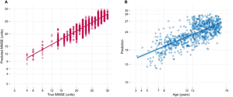
BackgroundThe retrogenesis hypothesis (RH) suggests that the functional and cognitive decline observed in Alzheimer's disease dementia mirrors in reverse order the brain development during childhood and adolescence.ObjectiveEquivalent electroencephalogram (EEG) patterns between older adults across different cognitive decline stages and children across different brain maturation stages were directly compared.MethodsTo capture the complex patterns that allow for such a comparison, a regression model was trained on EEG data from N = 510 older adults, at different stages of cognitive reserve, to identify EEG markers predictive of global cognitive status. The model was then applied on the same EEG markers of N = 696 children across different ages.ResultsThe model predicted MMSE scores with an average error of 2.53 and R2 of 0.80. When applied to children, predictions correlated positively with age (r = 0.73). Key predictors of cognitive function concordant in both populations were theta coherence (right frontal-left temporal/parietal), temporal Hjorth complexity, and beta edge frequency, supporting the RH.ConclusionsThese EEG features were inversely associated between older adults and children, supporting a functional underpinning of the retrogenesis model of dementia. Clinical validation of these biomarkers could favor their use in the continuous monitoring of cognitive function.

Keywords: Alzheimer's disease; Hjorth complexity; Mini-Mental State Exam; cognitive function; developmental age; electroencephalography; frontotemporal dementia; machine learning; spectral coherence.


Conflict of interest statement

Declaration of conflicting interestsThe authors declared the following potential conflicts of interest with respect to the research, authorship, and/or publication of this article: S.T. was member of advisory boards for Lilly, Eisai, Biogen, and GE Healthcare. He is member of the Independent Data Safety and Monitoring Board of the study ENVISION (Biogen). H.H. is an employee of Eisai Inc.; however, this article does not represent the opinion of Eisai. H.H. declares no competing financial interests related to the present article, and his contribution to this article reflects only and exclusively his academic and scientific expertise as part of an academic appointment at Sorbonne University, Paris, France. He serves as a Reviewing Editor and previously as Senior Associate Editor for the journal Alzheimer's & Dementia. Part of this study was initiated and developed in line with the Alzheimer's Precision Medicine Initiative (APMI) and Neurodegeneration Precision Medicine (NPMI) framework. The remaining authors declared no potential conflicts of interest with respect to the research, authorship, and/or publication of this article.

Figures

Figure 1.
Figure 1.
Regression of EEG features from older subjects (left, A) and of EEG features from children (right, B). A) Regression between predicted and true MMSE scores of validation examples of LOOCV procedure, with the older dataset. Each point represents one validation example independently evaluated on each CV fold. Regression is taken on all points (2297 points). B) Regression between the chronological age of children and the numerical predictions indicative of cognitive function outputted by the full-dataset MMSE regressor model. Each point represents one session of dataset P (789 points).
Figure 2.
Figure 2.
Accuracy of the numerical predictions in dataset P according to the known boundaries between MMSE and developmental age. A) Regression of Figure 1 where the predictions highlighted in green, when considered as MMSE scores, are coherent with the developmental ages proposed by the RH. B) Confusion matrix when considering a classification task with 3 counterpart groups of corresponding MMSE scores and developmental age intervals.
Figure 3.
Figure 3.
Most important features given by the trained model on the older dataset (left, A) and by the permutation testing on the children's dataset (right, B). On the left, feature importance is given by the mean decrease in impurity. On the right, feature importance is given by the non-decrease in error when permutating the respecting feature. The absolute values of both scales are not directly comparable. Highlighted in color are the features in common.
Figure 4.
Figure 4.
Partial dependence plots (PDPs) with highest positive (top panel) and negative (bottom panel) slope. In each PDP, the x-axis depicts the range of values each feature can take, and the y-axis shows the influence of each value the feature can take on the predicted outcome of the model, while holding all other features constant. The steepness of the slope indicates the strength of the feature's influence on the predicted outcome. All dataset P examples were evaluated on the trained full-dataset model.
Figure 5.
Figure 5.
Distribution of coherence (COH) features with significant predictive power for cognition over three MMSE and age counterpart groups. A) COH between right frontal and left temporal lobes in theta band; B) COH between right frontal and left parietal lobes in theta band. For each feature, all examples were normalized. Red is the distribution of the older population, and blue is the distribution of the children's population. Quartiles 1, 2 and 3 are drawn for the joint distributions of both populations. The KS statistic between each MMSE/age counterpart is given above each violin (* if KS p<0.05 ).
Figure 6.
Figure 6.
Topographic maps of Beta Edge Frequency (left, A) and Hjorth complexity (right, B), over two MMSE and age counterpart groups. For each feature, all examples were normalized. Cohort mean values are presented for the highlighted channels of interest. A) Channel O2 is highlighted, showing a positive correlation between its mean value and both MMSE and age. B) Channel T3 is highlighted, showing a negative correlation between its mean value and both MMSE and age.
Figure 7.
Figure 7.
Correlation maps between the most important features and target variables. A) Pair-wise Pearson correlation between the important features found in Figure 3A. B) Pair-wise Pearson correlation between the important features found in Figure 3B. In both matrices, in the lower triangle correlations were computed on the older dataset feature vectors, whereas in the upper triangle correlations were computed on the pediatric dataset feature vectors.

References

    1. Reisberg B, Franssen EH, Hasan SM, et al. Retrogenesis: clinical, physiologic, and pathologic mechanisms in brain aging, Alzheimer’s and other dementing processes. Eur Arch Psychiatry Clin Neurosci 1999; 249: S28–S36. - PubMed
    1. Reisberg B, Franssen EH, Souren LEM, et al. Evidence and mechanisms of retrogenesis in Alzheimer’s and other dementias: management and treatment import. Am J Alzheimers Dis Demen 2002; 17: 202–212. - PMC - PubMed
    1. Reisberg B, Ferris SH, Anand R, et al. Functional staging of dementia of the Alzheimer type. Ann N Y Acad Sci 1984; 435: 481–483.
    1. Xie C, Fong MC-M, Ma MK-H, et al. The retrogenesis of age-related decline in declarative and procedural memory. Front Psychol 2023; 14: 1212614. - PMC - PubMed
    1. Gayraud F, Loureiro IS, Frouin C, et al. Lexical deterioration in Alzheimer’s disease. In: 10th International Conference of Experimental Linguistics, Lisbonne, Portugal, pp. 101–104.

Publication types

LinkOut - more resources