. 2025 Jul;8(7):1337-1344.
doi: 10.1002/ame2.70052. Epub 2025 Jun 30.

Achieving scalable expansion of therapeutic porcine hepatocytes in vivo through serial transplantation

Affiliations

Achieving scalable expansion of therapeutic porcine hepatocytes in vivo through serial transplantation

Zhiqiang Han et al. Animal Model Exp Med. 2025 Jul.

Abstract

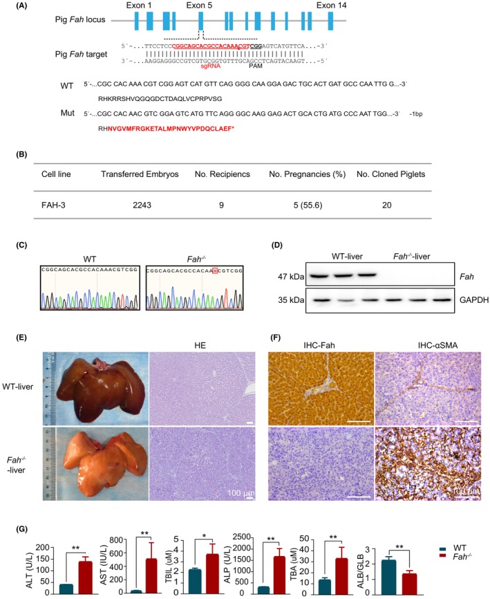
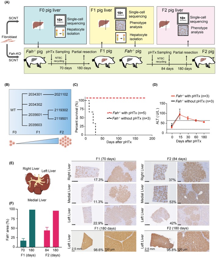
The clinical application of hepatocyte transplantation has been significantly hindered by the scarcity of primary hepatocytes and the functional immaturity of in vitro-produced hepatocytes. By performing serial allogeneic hepatocyte transplantation in CRISPR/Cas9-mediated Fah-knockout pigs, we successfully achieved large-scale expansion of hepatocytes while maintaining their authentic biological characteristics. Particularly, the established model enables sustained in vivo liver reconstruction, concurrently ameliorating hepatic fibrosis and demonstrating functional microenvironmental remodeling. Moreover, through comprehensive single-cell transcriptomic profiling of 52 418 hepatocytes across transplant generations (F0-F2), we discovered that the cellular composition of these transplanted hepatocytes is similar to that of wild-type hepatocytes. The regenerated liver exhibits all six major hepatic cell types identical to the wild-type counterparts, with the characteristic lobular zonation patterns well preserved. Our research provides valuable insights into the large-scale expansion of physiologically functional hepatocytes in vivo without compromising their biological properties. This finding holds great promise for advancing the clinical application of human hepatocyte transplantation, potentially offering more effective treatment options for patients with liver diseases.

Keywords: cellular therapy; hepatocyte; large‐scale expansion; regeneration; single‐cell RNA sequencing.


Conflict of interest statement

All authors declare that they have no conflict of interest.

Figures

FIGURE 1
FIGURE 1
Construction of the Fah −/− pig model, with its associated liver injury phenotype. (A) Pig Fah gene knockout site and prediction of the amino acid sequences of model pig genotypes. Red amino acids signify incorrect residues in the sequence. (B) Fah‐deficient model pig embryo transfer information. (C) Comparative analysis of Sanger sequencing peak maps. (D) Detection of Fah protein in the liver tissue. (E) Liver appearance and pathological detection of liver tissue H&E (hematoxylin and eosin) staining after death. Scale bar: 100 μm. (F) Immunohistochemical analysis of liver Fah and alpha‐smooth muscle actin (α‐SMA). Scale bar: 100 μm. (G) Detection of hepatic function indices in peripheral blood.
FIGURE 2
FIGURE 2
Continuous transplantation of pig hepatocytes into model pigs to reconstruct the liver for two generations. (A) Schematic of the continuous transplantation of porcine hepatocytes for two generations. The pig with a gray‐colored liver represents the Fah knockout pig, whereas the red‐colored liver represents either the liver of an F0 pig or a reconstructed liver. (B) Schematic of the recipients and sources of hepatocytes in a serial transplantation assay involving pig hepatocytes. (C) Survival curves of pigs after experimental hepatocyte transplantation (excluding swine killed during the experiment). (D) Peripheral blood alanine aminotransferase (ALT) in pigs after hepatocyte transplantation. (E) The sampling locations in different liver lobes. The Fah expression in various liver lobes from all reconstructed Fah‐knockout pig models were examined using immunohistochemistry (IHC) during the continuous transplantation of porcine hepatocytes. Scale bar: 2.5 mm or 100 μm. (F) Quantification of Fah immunohistochemical staining in regenerated hepatic tissue post‐reconstruction.
FIGURE 3
FIGURE 3
The classification of cell types and the gene expression profiles of livers that were subjected to the serial transplantation. (A) Uniform manifold approximation and projection (UMAP) plot of the distribution of different cell types in serial hepatocyte transplantation. (B) Dot plots of the expression levels of representative marker genes for hepatocytes in each subtype. Gray to red represents low to high gene expression. The size of dots indicates the percentage of cells with gene expression greater than zero. (C) Heat map (left) showing the differentially expressed genes (DEG) of three hepatocyte subtypes. The DEGs were divided into 14 modules according to the overlap of the three subtypes. Heat map (right) showing the Gene Ontology (GO) terms and pathways of the modules. (D) Sankey diagram showing the number of each cell type and the different cell numbers for consecutive generations of liver transplantation. (E) Hematoxylin and eosin (H&E) and Sirius Red staining of liver after hepatocyte transplantation. Scale bar: 500 μm. (F) The statistics of Sirius Red staining of fibrosis area. (G) Marker staining was conducted on a subset of cells of livers from various experimental groups. Scale bar: 50 μm. (H) Staining of the liver sections from the experimental liver samples. Scale bar: 50 μm.

References

    1. Zhang K, Zhang L, Liu W, et al. In vitro expansion of primary human hepatocytes with efficient liver repopulation capacity. Cell Stem Cell. 2018;23:806‐819.e804. doi: 10.1016/j.stem.2018.10.018 - DOI - PubMed
    1. Dhawan A, Puppi J, Hughes RD, Mitry RR. Human hepatocyte transplantation: current experience and future challenges. Nat Rev Gastroenterol Hepatol. 2010;7:288‐298. doi: 10.1038/nrgastro.2010.44 - DOI - PubMed
    1. Vons C, Beaudoin S, Helmy N, Dagher I, Weber A, Franco D. First description of the surgical anatomy of the cynomolgus monkey liver. Am J Primatol. 2009;71:400‐408. doi: 10.1002/ajp.20667 - DOI - PubMed
    1. Michalopoulos GK. Liver regeneration. J Cell Physiol. 2007;213:286‐300. doi: 10.1002/jcp.21172 - DOI - PMC - PubMed
    1. Azuma H, Paulk N, Ranade A, et al. Robust expansion of human hepatocytes in fah−/−/Rag2−/−/Il2rg−/− mice. Nat Biotechnol. 2007;25:903‐910. doi: 10.1038/nbt1326 - DOI - PMC - PubMed

LinkOut - more resources