. 2025 Aug 5;13(8):e0180624.
doi: 10.1128/spectrum.01806-24. Epub 2025 Jun 30.

Identification of relevant analytical methods for adeno-associated virus stability assessment during formulation development

Affiliations

Identification of relevant analytical methods for adeno-associated virus stability assessment during formulation development

Carina Rodenstein et al. Microbiol Spectr. .

Abstract

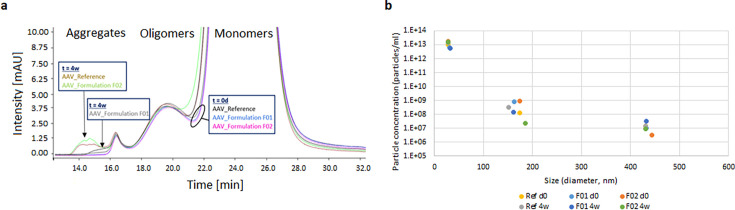
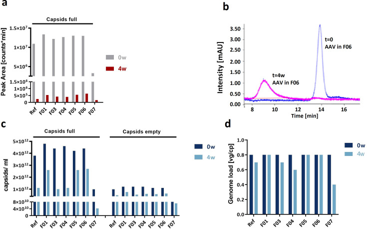
Formulation development for gene therapy viral vectors requires high-performance analytical methods that indicate or predict virus stability. Additionally, if testing involves multiple excipients, sample throughput must be sufficiently high, and the method should require low amounts of virus material. These are challenging and sometimes conflicting requirements, especially in the analysis of adeno-associated viral vectors, which are currently the most prominent viral vectors in gene therapy development. In this exploratory study, we evaluated the ability of different analytical methods to indicate or predict the stability of the adeno-associated viral vectors and identified highly suitable techniques that fulfilled the requirements for analysis during formulation development. As functionality assays, such as reporter gene assays, are already established for rapid quality control assessments, they are not addressed in this study. From this study, we conclude that two methods have great potential to predict the stability of adeno-associated viral vectors: extrinsic differential scanning fluorimetry using SYBR Gold as a DNA binding fluorescent dye to detect genome release and nano differential scanning fluorimetry to investigate capsid unfolding behavior (based on intrinsic protein fluorescence). Size-exclusion chromatography, using multi-angle light scattering and UV detection, was found to be stability-indicating for particle size distribution and determining genome load of adeno-associated viral vectors.IMPORTANCEViral vectors are of growing importance in the areas of gene therapy, oncology, and vaccine development. However, these vectors are very unstable and usually have to be stored frozen at very low temperatures due to sub-optimal formulation conditions, in many cases. The development of superior formulations for viral vectors requires high-performance analytical methods. In this study, we evaluated relevant analytical methods with respect to sample throughput, material consumption, and applicability for viral vector formulation development. To our knowledge, this is the first time that methods for viral vector analysis were categorized according to their power to predict or indicate time-dependent long-term stability. This categorization of analytical methods is essential to rationalize, accelerate, and enhance the formulation development of viral vectors. Therefore, the studies in this article are prerequisites for the development of more stable viral vectors for gene therapy and vaccines and higher yields in manufacturing.

Keywords: HPLC; adeno-associated virus; differential scanning calorimetry; fluorescence spectroscopy; forced conditions; formulation; gene delivery; light scattering (dynamic/static); stability.


Conflict of interest statement

The authors are either employees of Leukocare AG (Carina Rodenstein, Eva Schmid, Andreas Seidl) or Malvern Panalytical Ltd. (Natalia Marcova). There are no other conflicts of interest.

Figures

Fig 1
Fig 1
Analytical methods evaluated in this study.
Fig 2
Fig 2
SEC-UV at 280 nm (a) and MADLS (b) results of AAV-5 in reference buffer and formulation F01 and F02. For both techniques (a and b), the results of the measurement before (t = 0 days) and after (t = 4 weeks) thermal stress at 40°C for 4 weeks are depicted (n = 1). The arrows show additional aggregate formation for specific AAV-5 samples. For MADLS, encircled are the three detected populations of specific size.
Fig 3
Fig 3
Isothermal DLS results of AAV-5 in formulation F02 before (t = 0 days, blue) and after (t = 4 weeks, orange) thermal stress at 40°C for 4 weeks. Shown are the correlation curve (a) and the size distribution blotted against signal intensity (b) and volume percent (c).
Fig 4
Fig 4
DLS results of AAV-5 in the reference buffer and formulation F02 during thermal ramp application. Depicted are the samples from the accelerated aging study, AAV-5 in reference buffer before (a, t = 0 days) and after (b, t = 4 weeks) storage at 40°C for 4 weeks, and AAV-5 in formulation F02 before (c, t = 0 days) and after (d, t = 4 weeks) storage at 40°C for 4 weeks. Orange, AAV-monomer peak size; blue, overall scattering intensity is given in derived count rate.
Fig 5
Fig 5
Capsid protein unfolding (a) and DNA release (b) of AAV-2 in reference buffer and seven other formulations (F01–F07) measured by NanoDSF and eDSF (SYBR Gold), respectively. (a) NanoDSF (n = 2) of AAV-2 before (t = 0 days) and after (t = 4 weeks) thermal stress at 40°C for 4 weeks. Depicted are the parameters TON and Tm. (b) eDSF using SYBR Gold (n = 2) of AAV-2 before (t = 0 days) thermal stress at 40°C for 4 weeks. Depicted are the parameters TON and Tm. Samples for which no value was obtained are marked with *.
Fig 6
Fig 6
Thermogram as recorded by DSC of AAV-5 in reference buffer (a) and formulation F01 (b) before and after one freeze-thaw cycle.
Fig 7
Fig 7
Amount of full capsids for AAV-2 in seven different buffers before (t = 0 days) and after (t = 4 weeks) storage at 40°C for 4 weeks measured by AEX-HPLC (a and b) and SEC-MALS (c and d). (a) Relative area for full capsids determined by UV detection, (b) chromatogram of AAV-2 in formulation F06 measured at 260 nm, (c) concentration of full and empty capsids determined by SEC-MALS (UV-MALS detection), and (d) genome load depicted as ratio of full/empty capsids determined by SEC-MALS (UV-MALS detection).
Fig 8
Fig 8
AUC results of AAV-5 in reference buffer and formulations F01 and F02 before (t = 0 days, purple) and after storage at 40°C for 6 weeks (t = 6 weeks, blue). Capsid description: E (empty), PF (partially filled), and F (full). Others: HMWS (high-molecular-weight species) and LMWS (low-molecular-weight species).
Fig 9
Fig 9
Laser force cytology results for AAV-2 (a) and adenovirus (b). (a) The average velocity (left) and optical force index for AAV-2-infected and non-infected (NC) HEK-293 cells. (b) The correlation of adenovirus amount used for infection (multiplicity of infection, MOI) and the LFC parameter optical force index.
Fig 10
Fig 10
AAV attributes and analytical methods evaluated in this study for applicability and performance in AAV formulation development. Analytical methods in bold fulfilled the requirements for AAV formulation development.

References

    1. Horowitz ED, Rahman KS, Bower BD, Dismuke DJ, Falvo MR, Griffith JD, Harvey SC, Asokan A. 2013. Biophysical and ultrastructural characterization of adeno-associated virus capsid uncoating and genome release. J Virol 87:2994–3002. doi: 10.1128/JVI.03017-12 - DOI - PMC - PubMed
    1. Rayaprolu V, Kruse S, Kant R, Venkatakrishnan B, Movahed N, Brooke D, Lins B, Bennett A, Potter T, McKenna R, Agbandje-McKenna M, Bothner B. 2013. Comparative analysis of adeno-associated virus capsid stability and dynamics. J Virol 87:13150–13160. doi: 10.1128/JVI.01415-13 - DOI - PMC - PubMed
    1. Bennett A, Patel S, Mietzsch M, Jose A, Lins-Austin B, Yu JC, Bothner B, McKenna R, Agbandje-McKenna M. 2017. Thermal stability as a determinant of AAV serotype identity. Mol Ther Methods Clin Dev 6:171–182. doi: 10.1016/j.omtm.2017.07.003 - DOI - PMC - PubMed
    1. Burnham B, Nass S, Kong E, Mattingly M, Woodcock D, Song A, Wadsworth S, Cheng SH, Scaria A, O’Riordan CR. 2015. Analytical ultracentrifugation as an approach to characterize recombinant adeno-associated viral vectors. Hum Gene Ther Methods 26:228–242. doi: 10.1089/hgtb.2015.048 - DOI - PubMed
    1. Cole L, Fernandes D, Hussain MT, Kaszuba M, Stenson J, Markova N. 2021. Characterization of recombinant adeno-associated viruses (rAAVs) for gene therapy using orthogonal techniques. Pharmaceutics 13:586. doi: 10.3390/pharmaceutics13040586 - DOI - PMC - PubMed

LinkOut - more resources