. 2025 Jul 10;16(27):6935-6945.
doi: 10.1021/acs.jpclett.5c01189. Epub 2025 Jun 30.

Anomalous Water Fluorescence Induced by Solutes

Affiliations

Anomalous Water Fluorescence Induced by Solutes

Anna Maria Villa et al. J Phys Chem Lett. .

Abstract

The origin of recently reported anomalous fluorescence emissions from aqueous solutions of nonaromatic solutes remains elusive. To determine whether the solute nature influences the fluorescence characteristics and to identify a potential common mechanism, we measured the fluorescence spectra of 21 different solutions. We observed similar emission characteristics across all samples, suggesting that the solute nature plays a minimal role in the emission mechanism. Using time-dependent density functional theory on large water, NaCl/water, and glycerol/water clusters, we attributed the anomalous emission to the decay of charge-transfer-to-solvent excitations (CTTS) which populate a diradical zwitterionic excited state localized at hydrogen-bond network defects. The Arrhenius-like plots for NaCl and glycerol solutions revealed that the S1 nonradiative decay pathway involves the diradical recombination via librational motion. We propose that the presence of solute molecules slows this process, thus increasing the lifetime of the CTTS excited states and facilitating emission.


Figures

1
1
Comprehensive scatter plot displays the wavelength values for the maxima of the excitation and emission bands of all the aqueous solutions considered in this paper. The emission maxima cluster in three spectral regions centered at 295, 343, and 420 nm, while the excitation maxima cluster in two regions centered at 228 and 323 nm.
2
2
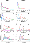
(A) Absorption spectra of NaCl aqueous solutions at different concentrations (4 M, 2 M, 1 M, 0.5 M, 0.25 M), the absorption spectrum of water is reported for comparison (light blue); (B) excitation and emission spectra of 1 M NaCl solution (excitation: blue = emission at 300 nm, light blue = emission at 350 nm; emission: red = excitation at 227 nm, green = excitation at 240 nm); (C) fluorescence emission spectra of NaCl aqueous solutions at different concentrations (4 M, 2 M, 1 M, 0.5 M, 0.25 M); excitation at 227 nm; (D) fluorescence emission spectra of 1 M NaCl aqueous solution at different temperatures; the emission spectra are recorded every 5 °C from 20 to 95 °C; excitation at 227 nm; (E) absorption spectra of glycerol aqueous solutions at different concentrations (4 M, 2 M, 1 M, 0.5 M, 0.25 M); (F) excitation and emission spectra of 1 M glycerol solution (excitation: light blue = emission at 305 nm, blue = emission at 410 nm; emission: red = excitation at 220 nm, green = excitation at 280 nm); (G) Fluorescence emission spectra of glycerol solutions at different concentrations (4 M, 2 M, 1 M, 0.5 M, 0.25 M, 0.125 M); excitation at 220 nm; (H) fluorescence emission spectra of 1 M glycerol aqueous solution at different temperatures; the emission spectra are recorded every 5 °C from 20 to 95 °C with excitation at 220 nm. Conditions: photomultiplier gain in B–D, F: 900 V; in G–H 770 V. Representative spectra from three independent experiments are shown.
3
3
At the top of the figure: the stability of the cluster model is considered with respect to the lowest energy structure identified. ΔE is the B-LYP/TZVP energy difference in kcal/mol. In the case of the glycerol water clusters, in blue and orange we report the solvent separated-like (SIP-like) and contact-pair like (CIP-like) forms in which one or more water molecules are or are not interposed between the two glycerol molecules. In the central part of the figure we report the structures for the most stable form (ΔE = 0 kcal/mol). The oxygen atoms and the hydrogen atoms involved in the CTTS band are evidenced in blue and red, respectively. In the bottom the HOMO and LUMO orbitals are reported for the most stable geometry form.
4
4
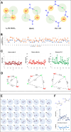
(A) The three (NaCl)2(H2O)108 cluster forms considered in the minimum energy search: contact ion pairs (C2v-like (NaCl)2), two CIP pairs separated by water (2­(NaCl)), and solvent-shared ion pairs (2Na+ + 2Cl). (B) Na+ and Cl coordination numbers (threshold 4 Å). (C) Average ion–ion distances (Å). (D) Lowest-energy structures of each type. (E) Sketches of 40 (Gly)2(H2O)108 isomers, highlighting glycerol–-glycerol H-bonds. (F) C2–C2 distances vs number of H-bonds, sorted by stabilization energy.
5
5
Scheme of CTTS S1 excitation and radiationless decay to S0 for (H2O)110 (A, blue spheres) and (NaCl)2(H2O)108 and (Gly)2(H2O)108 model clusters (B, green and yellow sphere, respectively). Dotted red PES represent the S n excited PES with n > 1. After light absorption, a S n excited state is populated and undergoes vibrational cooling decay to S1, passing through S n → S n‑1 intermolecular internal conversion (IC). For water clusters (A), S1 is characterized by an HOMO → LUMO CT transition localized at the cluster surface, with the formation of a zwitterionic diradical H2O•+/H2O•– solvated species. This latter evolves by vibrational cooling until reaching S1 → S0 IC. At this point, the solvated H2O•+/H2O evolve to a H3O+/OH ion-radical contact. On the S0 PES, this final species evolves rapidly by charge/radical recombination to the initial minimum geometry. In the case of glycerol (in light yellow) the CTTS state involves an HOMO → LUMO CT transition that is localized at the cluster surface, with the formation of a H2O•+/H2O•‑ or Gly•+/H2O•– solvated species; in the case of NaCl (in light green) TDDFT shows the formation of H2O•+/Na or Cl•/Na solvated species.

Similar articles

References

    1. Mallamace F.. The Liquid Water Polymorphism. Proc. Natl. Acad. Sci. U. S. A. 2009;106(36):15097–15098. doi: 10.1073/pnas.0908198106. - DOI - PMC - PubMed
    1. Hassanali A., Giberti F., Cuny J., Kühne T. D., Parrinello M.. Proton Transfer through the Water Gossamer. Proc. Natl. Acad. Sci. U. S. A. 2013;110(34):13723–13728. doi: 10.1073/pnas.1306642110. - DOI - PMC - PubMed
    1. Ceriotti M., Cuny J., Parrinello M., Manolopoulos D. E.. Nuclear Quantum Effects and Hydrogen Bond Fluctuations in Water. Proc. Natl. Acad. Sci. U. S. A. 2013;110(39):15591–15596. doi: 10.1073/pnas.1308560110. - DOI - PMC - PubMed
    1. Nilsson A., Pettersson L. G. M.. The Structural Origin of Anomalous Properties of Liquid Water. Nat. Commun. 2015;6:8998. doi: 10.1038/ncomms9998. - DOI - PMC - PubMed
    1. Huang C., Wikfeldt K. T., Tokushima T., Nordlund D., Harada Y., Bergmann U., Niebuhr M., Weiss T. M., Horikawa Y., Leetmaa M., Ljungberg M. P., Takahashi O., Lenz A., Ojamäe L., Lyubartsev A. P., Shin S., Pettersson L. G. M., Nilsson A.. The Inhomogeneous Structure of Water at Ambient Conditions. Proc. Natl. Acad. Sci. U. S. A. 2009;106(36):15214–15218. doi: 10.1073/pnas.0904743106. - DOI - PMC - PubMed

LinkOut - more resources