. 2025 Sep;49(9):1184-1196.
doi: 10.1002/cbin.70048. Epub 2025 Jun 30.

SIRT7-Mediated MVP Desuccinylation Facilitates Tongue Squamous Cell Carcinoma Progression by Activating JAK2/STAT3 Pathway

Affiliations

SIRT7-Mediated MVP Desuccinylation Facilitates Tongue Squamous Cell Carcinoma Progression by Activating JAK2/STAT3 Pathway

Zhuo Zhang et al. Cell Biol Int. 2025 Sep.

Abstract

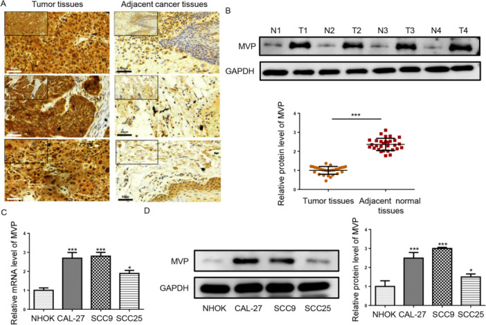
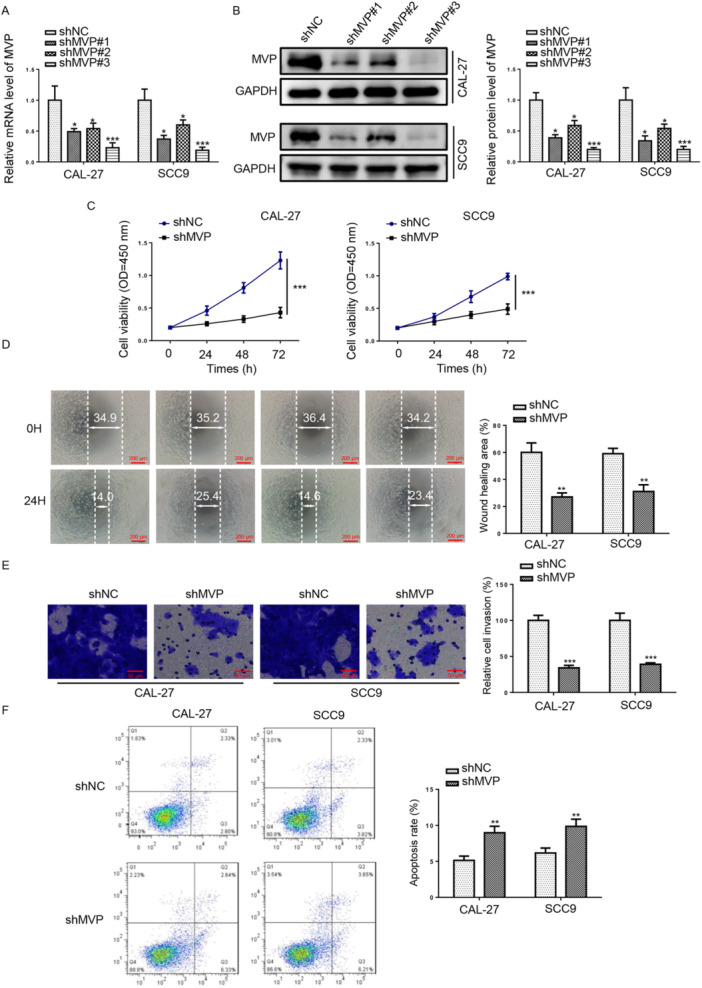
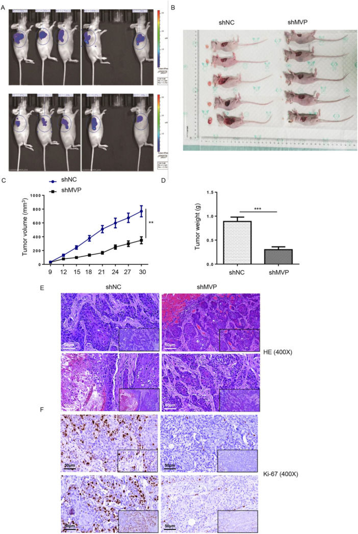
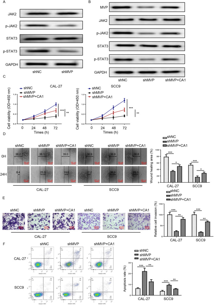
Major vault protein (MVP) plays a contributing role in multifarious cancers, and then its role in Tongue squamous cell carcinoma (TSCC) is uncomprehending. This study aimed to investigate the regulatory effect of MVP on malignant behavior of TSCC cells and its mechanism. We first pointed out the abnormal upregulation of MVP in tumor tissues by immunohistochemistry, western blot, and reverse transcription-quantitative polymerase chain reaction assays. Depletion of MVP hindered TSCC cell viability, migration, and invasion and accelerated apoptosis. Mechanistically, depletion of MVP inactivated Janus Kinase 2 (JAK2)/Signal Transducer and Activator of Transcription 3 (STAT3) pathway. Coumermycin A1 (CA1), a JAK2 agonist, was used to trigger JAK2/STAT3 signaling. Functional experiments demonstrated that CA1 significantly counteracted the inhibitory effects of MVP silencing on cell proliferation, invasion, and migration, as well as the stimulatory effects of MVP silencing on cell apoptosis. Moreover, we discovered that MVP undergoes succinylation and identified Sirtuin 7 (SIRT7) as the desuccinylase for MVP. Addition of SIRT7 promoted the protein stability of MVP in TSCC cells. Further, addition of MVP expedited the viability, migration, and invasion and suppressed apoptosis of TSCC cells, which was partly neutralized following depleted SIRT7. Our findings revealed that MVP desuccinylated by SIRT7 accelerated TSCC progression via regulating JAK2/STAT3 signaling.

Keywords: JAK2/STAT3 pathway; MVP; SIRT7; progression; tongue squamous cell carcinoma.


Figures

Figure 1
Figure 1
MVP expression was elevated in TSCC tissues and cells. (A) IHC (scale bars = 50 μm) was used to detect MVP expression in TSCC samples (n = 30) and adjacent normal tissues (n = 30). (B and C) RT‐qPCR and Western blot assays were performed to measure MVP mRNA and protein levels in TSCC tissues. (D and E) RT‐qPCR and Western blot assays were performed to measure MVP mRNA and protein levels in TSCC cells (CAL‐27, SCC9, and SCC25) and NHOK cells. *p < 0.05; **p < 0.01; ***p < 0.001.
Figure 2
Figure 2
Inhibition of MVP expression impeded TSCC cell malignant behaviors. (A and B) RT‐qPCR and Western blot assays were used to detect MVP mRNA and protein levels in TSCC cells transfected with sh‐NC, sh‐MVP#1, sh‐MVP#2, or sh‐MVP#3. (C) CCK‐8 assay was performed to measure cell viability of TSCC cells transfected with shNC or sh‐MVP. (D and E) Wound healing (scale bars = 200 μm) and transwell assays (scale bars = 50 μm) were used to measure cell migration and invasion in TSCC cells. (F) Cell apoptosis and cell cycle were assessed by flow cytometry assay. *p < 0.05; **p < 0.01; ***p < 0.001.
Figure 3
Figure 3
MVP promoted TSCC growth in vivo. (A) In vivo imaging was performed to evaluate the tumor burden in mice of sh‐MVP and sh‐NC groups. (B) Tumors were removed for collecting photos and weighing (C). (D) The volume of tumors formed in mice was measured and calculated at indicated time intervals. (E and F) HE and ki‐67 staining of tumors (scale bars = 50 μm). *p < 0.05; **p < 0.01; ***p < 0.001.
Figure 4
Figure 4
Depletion of MVP suppressed the activation of JAK2/STAT3 pathway in TSCC. (A) p‐JAK2, p‐STAT3, JAK2, and STAT3 protein levels were measured by western blot assay in TSCC cells transfected with shNC or sh‐MVP. The experiment was divided into the following groups: sh‐NC, sh‐MVP, sh‐MVP + CA1. (B) p‐JAK2, p‐STAT3, JAK2, and STAT3 protein levels were measured by western blot assay in TSCC cells. (C) Cell proliferation, (D) migration (scale bars=200μm), (E) invasion (scale bars = 50 μm), and (F) apoptosis were measured by CCK‐8, wound healing, transwell, and flow cytometry assays in TSCC cells. *p < 0.05; **p < 0.01; ***p < 0.001.
Figure 5
Figure 5
Depletion of SIRT7 enhanced MVP‐succ level in TSCC cells. (A and B) Proteins with binding potential to MVP were predicted on HitPredict and BIOGRID. (C) The interaction of MVP and SIRT7 was confirmed using co‐IP assay. (D) The succinylation level of MVP was detected in TSCC cells (CAL‐27, SCC9, and SCC25) by western blot. (E) The protein levels of MVP, SIRT7, and MVP‐ succinylation were measured by western blot in CAL‐27 and SCC9 cells. *p < 0.05; **p < 0.01; ***p < 0.001.
Figure 6
Figure 6
MVP was desuccinylated at K437 site. (A) The succinylation sites for MVP were predicted using the SuccinSite and GPSuc databases. (B) IP assay was performed to analyze the succinylation sites of MVP. (C) TSCC cells transfected with oe‐NC or oe‐SIRT7 vector were treated with CHX, then the protein expression of MVP was assayed by western blot at the different time points (0, 6, 12, and 24 h). (D) IF staining was utilized to assess the protein distribution of MVP and SIRT7 in TSCC cells (scale bars = 100 μm). *p < 0.05; **p < 0.01; ***p < 0.001.
Figure 7
Figure 7
Depletion of SIRT7 abolished TSCC cell malignant behaviors triggered by MVP. The experiment was divided into the following groups: oe‐NC, oe‐MVP, oe‐MVP+sh‐SIRT7. (A) MVP and SIRT7 protein levels were measured by western blot assay. (B) migration (scale bars = 200 μm), (C) invasion (scale bars = 50 μm), and (D) apoptosis were measured by CCK‐8, wound healing, transwell, and flow cytometry assays in TSCC cells. (E) p‐JAK2, p‐STAT3, JAK2, and STAT3 protein levels were measured by western blot assay in TSCC cells. *p < 0.05; ** p < 0.01; *** p < 0.001.

References

    1. Bray, F. , Ferlay J., Soerjomataram I., Siegel R. L., Torre L. A., and Jemal A.. 2018. “Global Cancer Statistics 2018: Globocan Estimates of Incidence and Mortality Worldwide for 36 Cancers in 185 Countries.” CA: A Cancer Journal for Clinicians 68, no. 6: 394–424. - PubMed
    1. Chen, X. F. , Tian M. X., Sun R. Q., et al. 2018. “Sirt 5 Inhibits Peroxisomal Acox 1 to Prevent Oxidative Damage and Is Downregulated in Liver Cancer.” EMBO Reports 19, no. 5: e45124. - PMC - PubMed
    1. Colella, G. , Rauso R., De Cicco D., et al. 2021. “Clinical Management of Squamous Cell Carcinoma of the Tongue: Patients Not Eligible for Free Flaps, a Systematic Review of the Literature.” Expert Review of Anticancer Therapy 21, no. 1: 9–22. - PubMed
    1. Colevas, A. D. , Yom S. S., Pfister D. G., et al. 2018. “Nccn Guidelines Insights: Head and Neck Cancers, Version 1.2018.” Journal of the National Comprehensive Cancer Network 16, no. 5: 479–490. - PubMed
    1. Deng, H. , Gong X., Ji G., Li C., and Cheng S.. 2023. “KIF2C Promotes Clear Cell Renal Cell Carcinoma Progression via Activating JAK2/STAT3 Signaling Pathway.” Molecular and Cellular Probes 72: 101938. - PubMed

LinkOut - more resources