. 2025 Jun 30;50(4):219.
doi: 10.1007/s11064-025-04473-0.

Propofol Modulates Microglial Glucose Metabolism Via the AMPK/HIF-1α Signaling Pathway To Ameliorate ECS-induced Cognitive Deficits in Depressive-like Rats

Affiliations

Propofol Modulates Microglial Glucose Metabolism Via the AMPK/HIF-1α Signaling Pathway To Ameliorate ECS-induced Cognitive Deficits in Depressive-like Rats

Mou Zou et al. Neurochem Res. .

Abstract

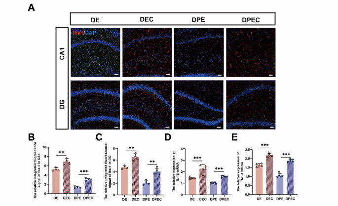
Propofol can partly ameliorate electroconvulsive shock (ECS)-induced learning and memory impairment by restoring synaptic plasticity. However, the exact mechanism is unknown. Microglia exert different immune functions by regulating their glucose metabolism, which is closely related to synaptic plasticity. We aimed to investigate whether the mechanism underlying the cognitive enhancement effects of propofol is associated with microglial glucose metabolism. Rats depression model was established by chronic unpredictable mild stress (CUMS). Sucrose preference test (SPT) and open field test (OFT) were used to detect anhedonia and anxiety-like behaviors in rats, respectively. Morris water maze (MWM) was used to evaluate the spatial learning and memory ability of rats. Transmission electron microscopy, immunofluorescence, enzymatic activity assays, Western blotting, and RT-qPCR were employed to evaluate hippocampal synaptic structural integrity, microglial glucose metabolism, and the expression of glycolytic regulators p-AMPK/AMPK and HIF-1α. The AMPK inhibitor compound C was used for reverse validation. Propofol attenuated the ECS-induced reduction of hippocampal synaptic proteins PSD-95 and SYN1, suppressed the upregulation of pro-inflammatory cytokines TNF-α and IL-1β, and reduced microglial activation. It also reduced the key glycolytic enzymes in microglia, increased AMPK expression, and decreased HIF-1α expression, thereby improving learning and memory impairment in ECS-treated rats. Compound C reversed propofol's neuroprotective effect. ECS-induced learning and memory deficits in depressive-like rats are associated with increased microglial glycolysis via the AMPK/HIF-1α pathway, a metabolism process that could be mitigated by propofol.

Keywords: Cognitive Impairment; Depression; Electroconvulsive Shock; Metabolic Reprogramming; Microglia; Propofol.


Conflict of interest statement

Declarations. Ethics Approval: All studies received ethical approval from the Ethics Committee of Chongqing Medical University (Chongqing, China). Competing Interests: The authors declare no competing interests.

Figures

Fig. 1
Fig. 1
Effect of electroconvulsive shock treatment on learning and memory functions in depressive-like rats. (A) Experimental timeline. (B) Sucrose preference percentage before and after ECS treatment. (C) The central activity time in the open field before and after ECS treatment. (D) Representative swimming trajectories. (E) Escape latency during the Morris water maze test. (F) Swimming speed. (G) Spatial exploration time in the target quadrant. n = 5 per group. (H) Transmission electron microscopy (magnification, 25,000×; scale, 500 nm) was used to observe the neural ultrastructure of the hippocampal region; in the figure, the presynaptic membrane is shown in yellow, the postsynaptic membrane is shown in purple; the circle indicates the intact postsynaptic membrane; the arrow points to discontinuous postsynaptic density. (I) Postsynaptic density thickness in the hippocampus. n = 5 per group. (J) The expression of synaptic proteins PSD-95 and SYN1 detected by western blot. (K–L) Semi-quantitative analysis of PSD-95 and SYN1. n = 3 per group. *p < 0.05, **p < 0.01, ***p < 0.001. ECS, electroconvulsive shock; PSD-95, postsynaptic density protein-95; SYN1, synapsin I
Fig. 2
Fig. 2
Temporal dynamics of hippocampal microglial glycolysis and activation in depressive-like rats following electroconvulsive shock. (A) Experimental timeline. (B) The mRNA expression of HK2, PFKFB3, and PKM in microglia detected by RT-qPCR. n = 4 per group. (C) Hexokinase activity, phosphofructokinase activity and pyruvate kinase activity in the hippocampus. n = 3 per group. (D) Immunofluorescence of Iba1 in the hippocampal CA1 and DG regions (magnification, 20 ×); relative fluorescence intensity was quantified in in the hippocampal CA1 and DG regions. n = 4 per group. (E) IL-1β expression detected by RT-qPCR. (F) TNF-α expression detected by RT-qPCR. (G) Arg-1 expression detected by RT-qPCR. (H) TGF-βexpression detected by RT-qPCR. n = 5 per group. ap < 0.05, compared with the control group; bp < 0.05, compared with DE1 group. HK2, hexokinase-2; PFKFB3, 6-phosphofructo-2-kinase; PKM, pyruvate kinase; Iba1, ionized calcium-binding adaptor molecule 1; TNF-α, tumor necrosis factor-alpha; IL-1β, interleukin 1-beta; Arg-1, arginase 1; RT-qPCR, real-time quantitative PCR
Fig. 3
Fig. 3
Effect of electroconvulsive shock treatment on learning and memory functions in depressive-like rats. (A) Experimental timeline. (B) Sucrose preference percentage before and after ECS treatment. (C) The central activity time in the open field before and after ECS treatment. (D) Representative swimming trajectories. (E) Escape latency during the Morris water maze test. (F) Spatial exploration time in the target quadrant. (G) Swimming speed. n = 5 per group. (H) Transmission electron microscopy (magnification, 25,000×; scale, 500 nm) was used to observe the neural ultrastructure of the hippocampal region; in the figure, the presynaptic membrane is shown in yellow, the postsynaptic membrane is shown in purple. (I) Postsynaptic density thickness in the hippocampus. n = 5 per group. (J) The expression of synaptic proteins PSD-95 and SYN1 detected by western blot. (K–L) Semi-quantitative analysis of PSD-95 and SYN1. n = 3 per group. *p < 0.05, **p < 0.01, ***p < 0.001. ECS, electroconvulsive shock; PSD-95, postsynaptic density protein-95; SYN1, synapsin I
Fig. 4
Fig. 4
Effect of propofol on microglial glycolysis in the hippocampus of depressive-like rats after electroconvulsive shock treatment. (A) The protein expression of p-AMPK, AMPK, and HIF-1α detected by western blot. (B, C) Semi-quantitative analysis of p-AMPK, AMPK, and HIF-1α. n = 3 per group. (D–F) The mRNA expression of HK2, PFKFB3, and PKM in microglia detected by real-time quantitative PCR. n = 5 per group. (G) Hexokinase activity in the hippocampus. (H) Phosphofructokinase activity in the hippocampus. (I) Pyruvate kinase activity in the hippocampus. n = 3 per group. (J) Double-labeling immunofluorescence of HK2 (green) and Iba1 (red) in the hippocampal CA1 region (magnification, 40 ×, scale bar = 40 μm); the relative number of HK2-positive microglia in the in the hippocampal CA1 region. n = 4 per group. *p < 0.05, **p < 0.01, ***p < 0.001. AMPK, AMP-activated protein kinase; HIF-1α, hypoxia-inducible factor 1-alpha; HK2, hexokinase-2; PFKFB3, 6-phosphofructo-2-kinase; PKM, pyruvate kinase; Iba1, ionized calcium binding adaptor molecule 1
Fig. 5
Fig. 5
Effect of propofol on microglial activation in the hippocampus of depressive-like rats after electroconvulsive shock treatment. (A) Immunofluorescence of Iba1 in the hippocampal CA1 and DG regions (magnification, 20 ×, scale bar = 50 μm). (B, C) Relative fluorescence intensity was quantified in the hippocampal CA1 and DG regions. n = 4 per group. (D) IL-1β expression detected by RT-qPCR. (E) TNF-α expression detected by RT-qPCR. n = 5 per group. *p < 0.05, **p < 0.01, ***p < 0.001. Iba1, ionized calcium-binding adaptor molecule 1; TNF-α, tumor necrosis factor-alpha; IL-1β, interleukin 1-beta; RT-qPCR, real-time quantitative PCR
Fig. 6
Fig. 6
Effect of propofol on microglial glycolysis in the hippocampus of depressive-like rats after electroconvulsive shock treatment. (A) Experimental timeline. (B) The protein expression of p-AMPK, AMPK, and HIF-1α detected by western blot. (C-D) Semi-quantitative analysis of p-AMPK, AMPK, and HIF-1α. n = 3 per group. (E) The mRNA expression of HK2 in microglia detected by real-time quantitative PCR. n = 5 per group. (F) Hexokinase activity in the hippocampus. (G) Phosphofructokinase activity in the hippocampus. (H) Pyruvate kinase activity in the hippocampus. n = 3 per group. (I) Double-labeling immunofluorescence of HK2 (green) and Iba1 (red) in the hippocampal CA1 region (magnification, 40 ×, scale bar = 40 μm); the relative number of HK2-positive microglia in the hippocampal CA1 region. n = 4 per group. *p < 0.05, **p < 0.01, ***p < 0.001. AMPK, AMP-activated protein kinase; HIF-1α, hypoxia-inducible factor 1-alpha; PFKFB3, 6-phosphofructo-2-kinase; HK2, hexokinase-2; Iba1, ionized calcium binding adaptor molecule 1
Fig. 7
Fig. 7
Effect of propofol on microglial activation in the hippocampus of depressive-like rats after electroconvulsive shock treatment. (A) Immunofluorescence of Iba1 in the hippocampal CA1 and DG regions (magnification, 20 ×, scale bar = 50 μm). (B, C) Relative fluorescence intensity was quantified in the hippocampal CA1 and DG regions. n = 4 per group. (D) IL-1β expression detected by RT-qPCR. (E) TNF-α expression detected by RT-qPCR. n = 5 per group. *p < 0.05, **p < 0.01, ***p < 0.001. Iba1, ionized calcium-binding adaptor molecule 1; TNF-α, tumor necrosis factor-alpha; IL-1β, interleukin 1-beta; RT-qPCR, real-time quantitative PCR
Fig. 8
Fig. 8
Effect of electroconvulsive shock treatment on learning and memory functions in depressive-like rats. (A) Sucrose preference percentage before and after ECS treatment. (B) The central activity time in the open field before and after ECS treatment. (C) Representative swimming trajectories. (D) Escape latency during the Morris water maze test. (E) Spatial exploration time in the target quadrant. (F) Swimming speed. n = 5 per group. (G) Transmission electron microscopy (magnification, 25,000×; scale, 500 nm) was used to observe the neural ultrastructure of the hippocampal region; in the figure, the presynaptic membrane is shown in yellow, the postsynaptic membrane is shown in purple. (H) Postsynaptic density thickness in the hippocampus. n = 5 per group. (I) The expression of synaptic proteins PSD-95 and SYN1 detected by western blot. (J–K) Semi-quantitative analysis of PSD-95 and SYN1. n = 3 per group. *p < 0.05, **p < 0.01, ***p < 0.001. ECS, electroconvulsive shock; PSD-95, postsynaptic density protein-95; SYN1, synapsin I
Fig. 9
Fig. 9
Mechanism diagram of propofol improving learning and memory function after ECS in depression-like rats by regulating glucose metabolism reprogramming of microglia through AMPK pathway

Similar articles

References

    1. Espinoza RT, Kellner CH (2022) Electroconvulsive Therapy[J]. N Engl J Med 386(7):667–672. 10.1056/NEJMra2034954 - PubMed
    1. Rose D, Fleischmann P, Wykes T, Leese M, Bindman J (2003) Patients’ perspectives on electroconvulsive therapy: systematic review[J]. BMJ (Clinical Res ed) 326(7403):1363. 10.1136/bmj.326.7403.1363 - PMC - PubMed
    1. Verwijk E, Comijs HC, Kok RM, Spaans HP, Stek ML, Scherder EJ (2012) Neurocognitive effects after brief pulse and ultrabrief pulse unilateral electroconvulsive therapy for major depression: a review[J]. J Affect Disord 140(3):233–243. 10.1016/j.jad.2012.02.024 - PubMed
    1. Bannerman DM, Sprengel R, Sanderson DJ, McHugh SB, Rawlins JN, Monyer H et al (2014) Hippocampal synaptic plasticity, Spatial memory and anxiety[J]. Nat Rev Neurosci 15(3):181–192. 10.1038/nrn3677 - PubMed
    1. Chen X, Nelson CD, Li X, Winters CA, Azzam R, Sousa AA et al (2011) PSD-95 is required to sustain the molecular organization of the postsynaptic density[J]. J Neuroscience: Official J Soc Neurosci 31(17):6329–6338. 10.1523/jneurosci.5968-10.2011 - PMC - PubMed

MeSH terms

LinkOut - more resources