Ultra-high-resolution and dual-energy computed tomography of carotid artery plaques differentiate symptomatic and asymptomatic patients by novel volumetric analysis
- PMID: 40587105
- PMCID: PMC12270255
- DOI: 10.1093/icvts/ivaf158
Ultra-high-resolution and dual-energy computed tomography of carotid artery plaques differentiate symptomatic and asymptomatic patients by novel volumetric analysis
Abstract
Objectives: The indication for carotid endarterectomy (CEA) mainly relies on the degree of stenosis and neurological symptoms. Plaque vulnerability has been associated with stroke risk, but identification on single-energy computed tomography (CT) has yielded heterogeneous results and is not routinely applied to clinical diagnostics. Hence, we intended to analyse CEA specimens for vulnerability features using dual-source CT and correlate these features with the presence of preprocedural symptoms.
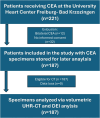
Methods: CT was performed on 187 carotid plaque specimens using ultra-high-resolution and dual-energy imaging on a dual-source scanner. Plaques were separated into calcified versus non-calcified volumes and analysed concerning HU-density, calcifications and volumetric dual-energy indices (DEIs). Comparative statistical analysis of plaque characteristics was performed with respect to the presence of neurological symptoms.
Results: The degree of stenosis of symptomatic and asymptomatic plaques was indifferent (69.2 ± 12.3% vs 66.3 ± 13.7%). The highest diagnostic accuracies were obtained by the % calcified volume (AUC 0.63 (0.54-0.71)), average whole plaque HU (AUC 0.71 (0.64-0.79)), profound calcification (AUC 0.74 (0.66-0.81)), calcification spots <1 mm (AUC 0.71 (0.63-0.79)) and spotty calcification (AUC 0.74 (0.66-0.82)). The diagnostic accuracy for symptomatic plaques was insignificant using average non-calcified plaque HU (AUC 0.59 (0.48-0.65)), but significant using average non-calcified plaque DEI (AUC 0.66 (0.58-0.74)).
Conclusions: Symptomatic plaques were identified best by measuring density of the whole, calcified or non-calcified plaque and via spotty, profoundly localized and less dense calcification. A volumetric DEI identifies symptomatic plaques with non-calcified plaque characteristics more accurately than single-energy CT. Future clinical studies are necessary to confirm these findings in patients.
Keywords: CT; carotid; plaque; stroke.
© The Author(s) 2025. Published by Oxford University Press on behalf of the European Association for Cardio-Thoracic Surgery.
Conflict of interest statement
J.T.: Funding by DFG (German Research Foundation)—TA1438/1-2.; speakers bureau Siemens Healthcare GmbH unrelated to this work; C.L.S. and F.B.: Siemens Healthineers, unrestricted research grant, speaker’s bureau; Others: no disclosures.
Figures


Similar articles
-
Differentiation of Atherosclerotic Carotid Plaque Components With Dual-Energy Computed Tomography.Invest Radiol. 2025 Aug 1;60(8):508-516. doi: 10.1097/RLI.0000000000001153. Epub 2025 Jan 22. Invest Radiol. 2025. PMID: 39836610
-
Ultrasound Characteristics of Symptomatic Carotid Plaques: A Systematic Review and Meta-Analysis.Cerebrovasc Dis. 2015;40(3-4):165-74. doi: 10.1159/000437339. Epub 2015 Aug 13. Cerebrovasc Dis. 2015. PMID: 26279159
-
Carotid Artery Surgery.2025 May 2. In: StatPearls [Internet]. Treasure Island (FL): StatPearls Publishing; 2025 Jan–. 2025 May 2. In: StatPearls [Internet]. Treasure Island (FL): StatPearls Publishing; 2025 Jan–. PMID: 28722976 Free Books & Documents.
-
Association of Carotid Plaque Calcification Attenuation With Intraplaque Hemorrhage Volume: 3D-Segmentation Analysis.J Neuroimaging. 2025 Jul-Aug;35(4):e70071. doi: 10.1111/jon.70071. J Neuroimaging. 2025. PMID: 40658028
-
Duplex ultrasound for diagnosing symptomatic carotid stenosis in the extracranial segments.Cochrane Database Syst Rev. 2022 Jul 11;7(7):CD013172. doi: 10.1002/14651858.CD013172.pub2. Cochrane Database Syst Rev. 2022. PMID: 35815652 Free PMC article.
References
-
- Crichton SL, Bray BD, McKevitt C et al. Patient outcomes up to 15 years after stroke. J Neurol Neurosurg Psychiatry 2016;87:1091–8. - PubMed
-
- Naylor R, Rantner B, Ancetti S et al. European Society for Vascular Surgery (ESVS) 2023 clinical practice guidelines on the management of atherosclerotic carotid and vertebral artery disease. Eur J Vasc Endovasc Surg 2023;65:7–111. - PubMed
-
- Eckstein HH, Kühnl A, Berkefeld J et al. S3-Leitlinie zur Diagnostik, Therapie und Nachsorge der extracraniellen Carotisstenose. Version 2.1. 2020. AWMF-register nr. 004-028. https://register.awmf.org/de/leitlinien/detail/004-028
-
- Aboyans V, Ricco JB, Bartelink MEL et al. ; ESC Scientific Document Group. 2017 ESC guidelines on the diagnosis and treatment of peripheral arterial diseases. Eur Heart J 2018;39:763–816. - PubMed
MeSH terms
Grants and funding
LinkOut - more resources
Full Text Sources
Medical

