. 2026 Apr 1;21(4):1396-1408.
doi: 10.4103/NRR.NRR-D-24-01029. Epub 2025 Mar 25.

Mitophagy: A key regulator in the pathophysiology and treatment of spinal cord injury

Affiliations

Mitophagy: A key regulator in the pathophysiology and treatment of spinal cord injury

Qiuyang Gu et al. Neural Regen Res. .

Abstract

Mitophagy is closely associated with the pathogenesis of secondary spinal cord injury. Abnormal mitophagy may contribute significantly to secondary spinal cord injury, leading to the impaired production of adenosine triphosphate, ion imbalance, the excessive production of reactive oxygen species, neuroinflammation, and neuronal cell death. Therefore, maintaining an appropriate balance of mitophagy is crucial when treating spinal cord injury, as both excessive and insufficient mitophagy can impede recovery. In this review, we summarize the pathological changes associated with spinal cord injury, the mechanisms of mitophagy, and the direct and indirect relationships between mitophagy and spinal cord injury. We also consider therapeutic approaches that target mitophagy for the treatment of spinal cord injury, including ongoing clinical trials and other innovative therapies, such as use of stem cells, nanomaterials, and small molecule polymers. Finally, we highlight the current challenges facing this field and suggest potential directions for future research. The aim of our review is to provide a theoretical reference for future studies targeting mitophagy in the treatment of spinal cord injury.

Keywords: ATP production disorders; cell death; mitochondria; mitophagy; neuroinflammation; neuroprotection; oxidative stress; secondary injury; spinal cord injury; treatment.


Conflict of interest statement

Conflicts of interest: The authors declare that there is no conflict of interest.

Figures

Figure 1
Figure 1
Occurrence and development of spinal cord injury. Spinal cord injury typically undergoes three stages: primary injury, secondary injury, and chronic injury. Primary injury is often caused by various external factors. This subsequently triggers secondary injury, including blood vessel rupture, macrophage and microglial infiltration, inflammation, mitochondrial damage, and ROS accumulation. Chronic injury is characterized by glial scarring and prolonged neuroinflammation. CD86: Cluster of differentiation 86; IFN-γ: interferon gamma; IGF-CSF: insulin-like growth factor-colony stimulating factor; IL-1β: interleukin 1 beta; IL-6: interleukin 6; IL-12: interleukin 12; iNOS: inducible nitric oxide synthase; LPS: lipopolysaccharide; ROS: reactive oxygen species; TNF-α: tumor necrosis factor alpha.
Figure 2
Figure 2
Mechanisms of secondary spinal cord injury. A growing body of evidence indicates that mitochondrial dysfunction and abnormal mitophagy are important mechanisms underlying secondary SCI. These mechanisms directly or indirectly cause ATP production disorder, ROS overproduction, neuroinflammation, and nerve cell death. Additionally, abnormal mitophagy renders the physiological mechanism of regulating mitochondrial function and monitoring mitochondrial quality ineffective. Abnormal mitochondria cannot be timely cleaned up, causing continued damage to neuronal cells. As such, modulating neuronal mitophagy may be a practical approach to promote the recovery of neurological function after SCI. ATP: Adenosine triphosphate; IL-11: interleukin 11; PCD: programmed cell death; ROS: reactive oxygen species; TLR: Toll-like receptor; TNF-α: tumor necrosis factor α.
Figure 3
Figure 3
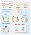
Overview of mitophagy. (A) Mechanisms of mitophagy. Mitophagy is activated by stressors including hypoxia, nutrient deficiency, DNA damage, inflammation, and mitochondrial membrane depolarization. It plays a role in maintaining mitochondrial integrity and function. The process of mitophagy encompasses initiation, membrane nucleation and phagosome formation and expansion, fusion with lysosomes, and ultimate degradation. (B) Main mitophagy pathways. Mitophagy is primarily achieved through two pathways, i.e., the ubiquitin-dependent pathway and the ubiquitin-independent pathway. The ubiquitin-dependent pathway relies on the ubiquitination of mitochondrial surface proteins to promote mitophagy, with the PINK1/Parkin pathway being the most extensively studied. The ubiquitin-independent pathway is majorly mediated by autophagy receptors, which directly bind to LC3 without ubiquitination, thereby initiating mitophagy. BCL2L13: Bcl2 like 13; BNIP3: BCL2 19 kDa interacting protein 3; FKBP8: FK506-binding protein 8; FUNDC1: FUN14 domain-containing protein 1; LC3: microtubule associated protein 1 light chain 3; NBR1: neighbor of BRCA1 gene 1; NDP52: nuclear dot protein 52 kDa; NIX: NIP3-like protein X; OMM: outer mitochondrial membrane; OPTN: optineurin; P62: sequestosome 1; PHB2: Prohibitin 2; PINK1: PTEN induced putative kinase 1; TAX1BP1: Tax1-binding protein 1; Ub: ubiquitin.
Figure 4
Figure 4
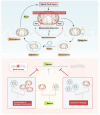
Summary of the mechanism of action of mitophagy in spinal cord injury. Mitochondria play an important role in spinal cord injury. After spinal cord injury, mitochondria undergo oxidative stress damage, leading to adverse outcomes, including neuronal injury and inflammatory infiltration. Appropriate mitophagy can rescue this damage and facilitate recovery from spinal cord injury. However, excessive or insufficient mitophagy will disrupt this balance, which is detrimental to the treatment of spinal cord injury. ATP: Adenosine triphosphate; ROS: reactive oxygen species.
Figure 5
Figure 5
Timeline of research progress in the role of mitophagy in acute central nervous system. BNIP3: BCL2 19 kDa interacting protein 3; GIT1: G-protein coupled receptor kinase interacting protein 1; LC3: microtubule associated protein 1 light chain 3; NIX: NIP3-like protein X; ROS: reactive oxygen species; SCI: spinal cord injury.

References

    1. Agrawal I, Jha S. Mitochondrial dysfunction and Alzheimer’s disease: role of microglia. Front Aging Neurosci. 2020;12:252. - PMC - PubMed
    1. Ahuja CS, Wilson JR, Nori S, Kotter MRN, Druschel C, Curt A, Fehlings MG. Traumatic spinal cord injury. Nat Rev Dis Primers. 2017;3:17018. - PubMed
    1. Ahuja CS, Nori S, Tetreault L, Wilson J, Kwon B, Harrop J, Choi D, Fehlings MG. Traumatic spinal cord injury-repair and regeneration. Neurosurgery. 2017;80:S9–S22. - PubMed
    1. Aimaiti M, Wumaier A, Aisa Y, Zhang Y, Xirepu X, Aibaidula Y, Lei X, Chen Q, Feng X, Mi N. Acteoside exerts neuroprotection effects in the model of Parkinson’s disease via inducing autophagy: network pharmacology and experimental study. Eur J Pharmacol. 2021;903:174136. - PubMed
    1. Ajoolabady A, Chiong M, Lavandero S, Klionsky DJ, Ren J. Mitophagy in cardiovascular diseases: molecular mechanisms, pathogenesis, and treatment. Trends Mol Med. 2022;28:836–849. - PMC - PubMed

LinkOut - more resources