. 2025 Jun 30;21(6):e1013256.
doi: 10.1371/journal.ppat.1013256. eCollection 2025 Jun.

Arabidopsis CNL receptor SUT1 confers immunity in hydathodes against the vascular pathogen Xanthomonas campestris pv. campestris

Affiliations

Arabidopsis CNL receptor SUT1 confers immunity in hydathodes against the vascular pathogen Xanthomonas campestris pv. campestris

Nanne W Taks et al. PLoS Pathog. .

Abstract

Bacterial plant pathogens exploit natural openings, such as pores or wounds, to enter the plant interior and cause disease. Plants guard these openings through defense mechanisms. However, bacteria from the genus Xanthomonas have specialized in that they enter their host via a special entry point, the hydathode-an organ at the leaf margin involved in xylem sap guttation. Hydathodes can mount an immune response against bacteria, including non-adapted and adapted pathogens like X. campestris pv. campestris (Xcc) that cause vascular disease. Previously, it was shown that the RKS1/ZAR1 immune complex confers vascular resistance against Xcc by recognizing XopAC activity, a type III effector (T3E). However, in absence of XopAC recognition, Arabidopsis Col-0 hydathodes still display resistance against Xcc. Here we mapped the causal gene using an inoculation method that promotes Xcc hydathode entry. Using a population of Recombinant Inbred Lines (RILs) of a cross between a susceptible (Oy-0) and resistant accession (Col-0), a major QTL for Xcc resistance was found on the right arm of Chromosome 5 in Col-0. Combining this result with a genome-wide association analysis yielded a single candidate gene encoding a coiled-coil nucleotide-binding leucine-rich repeat (CNL-type) immune receptor protein called SUPPRESSOR OF TOPP4 1 (SUT1). Expression of SUT1 was confirmed in hydathodes. We reveal that RKS1/ZAR1 and SUT1 confer different levels of Xcc resistance in different tissue types. Both RKS1/ZAR1 and SUT1 are alone sufficient for Xcc resistance in Col-0 hydathodes. However, RKS1/ZAR1 resistance is also effective in tissue types that represent late infection stages, i.e., xylem and mesophyll. In contrast, SUT1 resistance is not effective in the xylem, while weakly additive to RKS1/ZAR1 in the mesophyll. We thus identify a novel R gene, SUT1, that confers Xcc resistance primarily early in the infection during hydathode colonization.


Conflict of interest statement

I have read the journal's policy and the authors of this manuscript have the following competing interests: H.A.v.d.B. is currently an employee of Rijk Zwaan. This work was supported in part by a grant from Rijk Zwaan and Bejo Zaden. Both companies had no role in the design of the study, data collection, and writing of the manuscript.

Figures
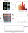
Fig 1
Fig 1. Mapping of SUT1 as a candidate R gene against Xcc8004 ΔxopAC in Arabidopsis Col-0.
A) Disease symptoms and bacterial luminescence in the resistant Col-0 and susceptible Oy-0 accessions following spray inoculation with bioluminescent Xcc8004 ΔxopAC. Leaves shown are representative of spray inoculation with either Tn5:lux or Tn7:lux tagged reporter strains. Bacterial spread was visualized using light-sensitive film 14 days post inoculation (dpi). B) Histogram depicting the variation in luminescence index score 14 dpi (normalized to Oy-0) for all 165 Oy-0 × Col-0 Recombinant Inbred Lines (RILs) following spray inoculation with Xcc8004 ΔxopAC Tn5:lux. C) Population structure of 165 RILs in the Oy-0 × Col-0 RIL population, ordered by luminescence index score at 14 dpi (normalized to Oy-0) shown in (B). D) QTL graph showing LOD scores for each of the 85 markers in the RIL population. LOD significance threshold determined by permutation (n = 1000) at 2.49 E) Manhattan plot of Chromosome 5 from a screen of 321 accessions present in the HapMap collection. Highlighted SNPs in orange are located within the coding sequence of SUT1. False Discovery Rate (FDR) thresholds set at -log10 (p) = 8 (Bonferroni) and -log10 (p) = 7.29 (Benjamini-Hochberg). See also S1 Fig for the full Manhattan plot.
Fig 2
Fig 2. SUT1 contributes to hydathode resistance against Xcc8004 ΔxopAC.
A) Predicted SUT1 protein model. Arrows depict predicted amino acid differences between Oy-0 vs. Col-0. B) SUT1 gene model depicting the Col-0 allele and approximate integration site of the sut1-8 and sut1-9 T-DNA insertions. C) Spray inoculation of sut1-8 and sut1-9 with Xcc8004 ΔxopAC Tn7:lux. Wildtype background (Col-0), bak1-5;bkk1-1 (Col-0) and the accession Oy-0 were included as controls. Top, visualization of high density hydathode colonization using light sensitive films (7 dpi); bottom, visualization of the systemic spread of Xcc and disease symptom development (14 dpi). D,E,F) Number of infected hydathodes 7 dpi (D), chlorotic area 14 dpi (E) and bacterial colonization 14 dpi (F) per leaf for each line shown in panel C following spray inoculation with Xcc8004 ΔxopAC Tn7:lux (n = 21 leaves per experiment). In panel D, the circle size depicts the number (#) of leaves with a certain score (y-axis) from three independent repetitions. Significance letters from non-parametric Kruskal-Wallis test with Dunn’s Post-Hoc test, p-value threshold = 0.05.
Fig 3
Fig 3. SUT1 is expressed in epithem cells within hydathodes.
A) Arabidopsis pSUT1::GUS lines stained with X-Gluc show SUT1 expression over the entire leaf (4-week-old plants). GUS staining is stronger in the hydathodes along the leaf margin (white arrows). B) Arabidopsis epithem marker line pPUP1::RCl2a-tdTomato shows tdTomato localization in epithem cell membrane within hydathodes. Epithem tissue can be distinguished from the mesophyll cells in the bright-field image (white dashed line). C) Arabidopsis pSUT1::NLS3 × mVENUS T2 line shows mVENUS accumulation in nuclei of mesophyll and epithem cells. Epithem cells can be distinguished from mesophyll cells in the bright-field image as a region with dense small cells on the leaf margin (white dashed line). Images were taken of epidermal peels and are representative of three independent lines (S5 Fig).
Fig 4
Fig 4. SUT1 resistance is compromised when the Xcc inoculation bypasses hydathode colonization.
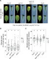
A) Disease symptoms and bacterial spread in Col-0, sut1-9, bak1-1;bkk1-5 and Oy-0 leaves at 7 dpi following clip inoculation with Xcc8004 ΔxopAC Tn7:lux. Bacterial progression is quantified as the spread of the luminescence signal along the midvein. B,C) Chlorotic leaf area (B) and bacterial spread along the midvein (C) 7 dpi following clip inoculation with Xcc8004 ΔxopAC Tn7:lux. SUT1 resistance against Xcc is apparently ineffective in the vasculature of Col-0 when the hydathodes are bypassed and the bacteria are introduced using leaf clipping. Significance letters from a two-way ANOVA with Tukey Post-Hoc test, p-value threshold = 0.05 (n = 12 leaves per experiment).
Fig 5
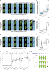
Fig 5. RKS1/ZAR1 and SUT1 confer resistance to Xcc in different stages of infection.
A) Spray inoculation with Xcc8004 Tn7:lux on rks1-1 and sut1-9 single mutants and two independent rks1/sut1 CRISPR knockout lines with wildtype background Col-0 and accession Oy-0 as controls. Top, visualization of Xcc hydathode colonization using light sensitive films (7 dpi); bottom, visualization of the systemic spread of Xcc and disease symptom development (14 dpi). B,C) Quantification of panel A as number of infected hydathodes per leaf at 7 dpi (B) and bacterial colonization (C) at 14 dpi (n = 18 leaves per experiment). D) Clip inoculation with Xcc8004 Tn7:lux on the same plant lines used in panel A. E) Quantification of bacterial progression along the midvein of panel D at 5 dpi (n = 18 leaves per experiment). F) Colony forming units at 0, 3 and 5 days post syringe inoculation of Xcc8004 WT Tn7:lux in the apoplast of the plant lines used in panel A (n = 8 samples per experiment. G) Schematic model indicating the contribution to resistance by RKS1/ZAR1 and SUT1 in different leaf tissues.

Similar articles

References

    1. Melotto M, Zhang L, Oblessuc PR, He SY. Stomatal defense a decade later. Plant Physiol. 2017;174(2):561–71. doi: 10.1104/pp.16.01853 - DOI - PMC - PubMed
    1. Zhang J, Coaker G, Zhou J-M, Dong X. Plant immune mechanisms: from reductionistic to holistic points of view. Mol Plant. 2020;13(10):1358–78. doi: 10.1016/j.molp.2020.09.007 - DOI - PMC - PubMed
    1. Snoeck S, Johanndrees O, Nürnberger T, Zipfel C. Plant pattern recognition receptors: from evolutionary insight to engineering. Nat Rev Genet. 2025;26(4):268–78. doi: 10.1038/s41576-024-00793-z - DOI - PubMed
    1. Liu Z, Hou S, Rodrigues O, Wang P, Luo D, Munemasa S, et al. Phytocytokine signalling reopens stomata in plant immunity and water loss. Nature. 2022;605(7909):332–9. doi: 10.1038/s41586-022-04684-3 - DOI - PMC - PubMed
    1. Melotto M, Underwood W, Koczan J, Nomura K, He SY. Plant stomata function in innate immunity against bacterial invasion. Cell. 2006;126(5):969–80. doi: 10.1016/j.cell.2006.06.054 - DOI - PubMed

MeSH terms

Substances