Review
. 2025 Jul;41(5):e70065.
doi: 10.1002/dmrr.70065.

Concomitant Diabetes and Atrial Fibrillation: Epicardial Fat and Macrophage-Related Mechanisms

Affiliations
Review

Concomitant Diabetes and Atrial Fibrillation: Epicardial Fat and Macrophage-Related Mechanisms

Saja Al-Rubaye et al. Diabetes Metab Res Rev. 2025 Jul.

Abstract

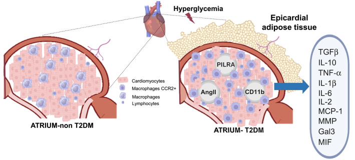
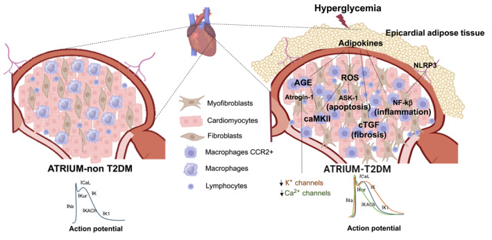
Type 2 diabetes mellitus (T2DM) is present in 25% of patients with atrial fibrillation (AF), the most prevalent arrhythmia in the world. This concomitant disorder enhances thromboembolic events, length of hospital stay after AF ablation, renal impairment after anticoagulation, heart rate variability after glucose-lowering treatment, and cardiac mortality. These patients accumulate inflamed epicardial fat (EAT) with paracrine consequences on β-oxidation of mitochondria, cytosolic Ca2+ fluxes, and sarcomere shortening. Knowing these specific targets will improve the efficacy of personalised preventive and curative therapies since AF leads to AF and EAT accumulation. This review tries to clarify the interplay among epicardial fat accumulation and macrophages with concomitant T2DM and AF to provide a summary of current known mechanisms and therapeutic strategies.

Keywords: atrial fibrillation; diabetes; epicardial fat; macrophages; mechanisms.


Conflict of interest statement

The authors declare no conflicts of interest.

Figures

FIGURE 1
FIGURE 1
Macrophages and T2DM in AF. Left atrium from the patient without type 2 diabetes mellitus (T2DM) (on left) and with T2DM (on right) with extracellular hyperglycemia, epicardial adipose tissue accumulation, cellular composition, and activation of several second messengers involved in pro‐inflammatory macrophage profile and inflammation. Hyperglycemia induces infiltration of macrophages by (a) high circulating levels of Paired Immunoglobulin‐Like Type 2 Receptor Alpha (PILRA), (b) monocytes‐CD11, and (c) Angiotensin II (AngII). Pro‐inflammatory macrophages can induce the secretion of several chemokines and cytokines that promote left atrial enlargement and structural remodelling.
FIGURE 2
FIGURE 2
Pathophysiological mechanisms of T2DM in AF. Left atrium from the patient without type 2 diabetes mellitus (T2DM) (left) and with T2DM (right) with extracellular hyperglycemia, epicardial adipose tissue accumulation, cellular composition, and activation of several second messengers involved in structural and electrical remodelling. Hyperglycemia induces (a) overactivity of the sympathetic system in the heart, (b) myocardial apoptotic signalling through activation of apoptosis signal‐regulating kinase 1 (Ask‐1), (c) fibrosis signalling with an increment of connective tissue growth factor (CTGF), (d) advanced glycation end products (AGE) and oxidative stress that induces abnormalities on calcium through calmodulin‐dependent protein kinase‐II (caMKII) activity, (e) inflammation through the NLRP3 inflammasome and NF‐kB pathway, and (f) changes on action potential duration through downregulation of potassium channels, degraded by the activity of Atrogin‐1.

Similar articles

References

    1. Palmer A. K. and Jensen M. D., “Metabolic Changes in Aging Humans: Current Evidence and Therapeutic Strategies,” Journal of Clinical Investigation 132, no. 16 (2022), 10.1172/jci158451. - DOI - PMC - PubMed
    1. Karadag B., Ozulu B., Ozturk F. Y., Oztekin E., Sener N., and Altuntas Y., “Comparison of Epicardial Adipose Tissue (EAT) Thickness and Anthropometric Measurements in Metabolic Syndrome (MS) Cases above and under the Age of 65,” Archives of Gerontology and Geriatrics 52, no. 2 (2011): e79–e84, 10.1016/j.archger.2010.06.016. - DOI - PubMed
    1. Song D. K., Hong Y. S., Lee H., Oh J. Y., Sung Y. A., and Kim Y., “Increased Epicardial Adipose Tissue Thickness in Type 2 Diabetes Mellitus and Obesity,” Diabetes & Metabolism Journal 39, no. 5 (2015): 405, 10.4093/dmj.2015.39.5.405. - DOI - PMC - PubMed
    1. Pavlou S., Lindsay J., Ingram R., Xu H., and Chen M., “Sustained High Glucose Exposure Sensitizes Macrophage Responses to Cytokine Stimuli but Reduces Their Phagocytic Activity,” BMC Immunology 19 (2018): 1–13, 10.1186/s12865-018-0261-0. - DOI - PMC - PubMed
    1. Santiago‐Fernández C., Pérez‐Belmonte L. M., Millán‐Gómez M., et al., “Overexpression of Scavenger Receptor and Infiltration of Macrophage in Epicardial Adipose Tissue of Patients With Ischemic Heart Disease and Diabetes,” Journal of Translational Medicine 17, no. 1 (December 2019): 95, 10.1186/s12967-019-1842-2. - DOI - PMC - PubMed

MeSH terms