. 2025 Sep:85:103742.
doi: 10.1016/j.redox.2025.103742. Epub 2025 Jun 26.

Evidence for alcohol-mediated hemolysis and erythrophagocytosis

Affiliations

Evidence for alcohol-mediated hemolysis and erythrophagocytosis

Chaowen Zheng et al. Redox Biol. 2025 Sep.

Abstract

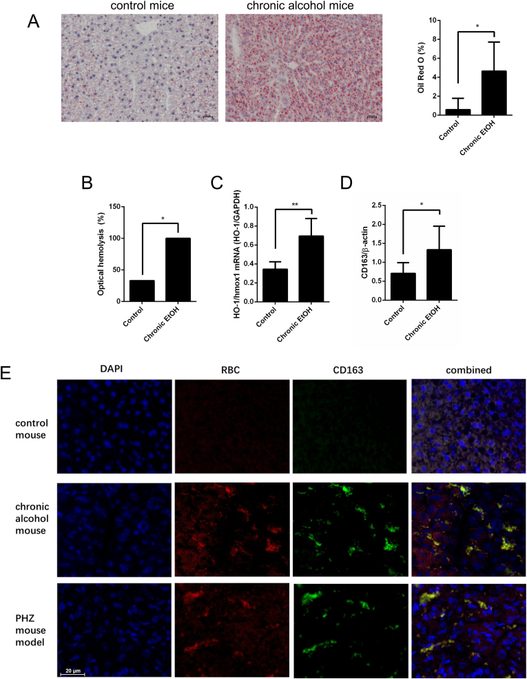
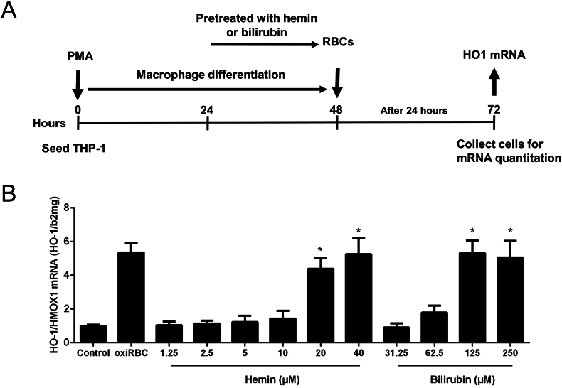
Alcohol-related liver disease (ALD) is the most common liver disease worldwide; however, its underlying molecular mechanisms remain poorly understood. Here, we identify ethanol-mediated hemolysis and erythrophagocytosis as major contributors to ALD pathogenesis using both in vitro and in vivo models, as well as surrogate markers such as heme oxygenase-1 (HO-1) and CD163, a scavenger receptor for hemoglobin-haptoglobin complexes. A key initial observation was the direct optical evidence of serum hemolysis in heavy drinkers, which diminished after one week of alcohol withdrawal. In parallel, soluble CD163 (sCD163) levels declined during alcohol detoxification correlating with liver damage and fibrosis stages. Moreover, red blood cells (RBCs) from heavy drinkers exhibited increased fragility under hemolytic stress. In ethanol-fed mice, we also observed serum hemolysis. Erythrophagocytosis in liver tissue was visualized by co-localization of CD163 and hemoglobin autofluorescence. In vitro studies confirmed that ethanol - at concentrations transiently present in the upper gastrointestinal tract during alcohol ingestion - directly induces hemolysis and primes RBCs for erythrophagocytosis through eryptosis, marked by externalization of phosphatidylserine. Both heme, released during hemolysis, and bilirubin, its degradation product, further amplified erythrophagocytosis at clinically relevant concentrations, suggesting a self-perpetuating cycle. The antioxidant N-acetylcysteine efficiently blocked ethanol-induced RBC priming for erythrophagocytosis. In conclusion, alcohol triggers a cascade of hemolysis, eryptosis, and erythrophagocytosis that may contribute to the pathogenesis of alcoholic hepatitis and end-stage ALD. sCD163 could serve as a noninvasive marker of hemolysis-associated macrophage activation. This mechanism opens new avenues for antioxidant-based therapies and may help to explain typical iron abnormalities, including ferroptosis, and hyperbilirubinemia in ALD.

Keywords: Alcohol-related liver disease; Alcoholic hepatitis; Bilirubin; CD163; Eryptosis; Erythrophagocytosis; Ferroptosis; Heme; Hemoglobin; Hemolysis; Iron overload; Liver cirrhosis; Red blood cell.


Conflict of interest statement

Declaration of competing interest The authors declare that they have no known competing financial interests or personal relationships that could have appeared to influence the work reported in this paper.

Figures

Image 1
Graphical abstract
Fig. 1
Fig. 1
Signs of hemolysis, enhanced heme turnover and increased RBC fragility in heavy drinkers. A) Example of macroscopic optical signs of hemolysis in a serum sample before and one week after alcohol detoxification. B) Significantly decreased signs of optical hemolysis determined by optical inspection in patient sera from heavy drinkers after alcohol withdrawal (see methods section). Frozen serum samples from n = 439 heavy drinkers prior and one week after alcohol detoxification were used. Of note, the data suggest that one week of alcohol detoxification already improves ethanol-mediated RBC fragility. C) Serum sCD163 levels (soluble hemoglobin-haptoglobin scavenging receptor) measured by ELISA before and one week after alcohol detoxification in n = 72 heavy drinkers. Shown is median and interquartile range. D and E) RBCs of heavy drinkers are more fragile in response to hemolytic stress by the hemolytic agent phenylhydrazine (PHZ) or mechanically during blood taking. RBCs from both heavy drinkers and healthy volunteers (each cohort n = 6) were treated in vitro with PHZ for 60 min and hemolysis was measured by absorption spectroscopy in the supernatant (see methods section). F) Direct exposure of human RBCs with ethanol (EtOH) for 24 h. Note, that hemolysis or modification only occurs at quite high levels of ethanol (ca. 10 %) which, however, may be achieved during binge drinking in some compartments using high percentage liquors or wine. Exposure time can be as short as 60min to lyse RBCs. ∗P < 0.05, ∗∗P < 0.01, ∗∗∗P < 0.001, ∗∗∗∗P < 0.0001.
Fig. 2
Fig. 2
CD163 mRNA expression in living precision cut liver slices (PCLS) treated with different RBC conditions. This figure shows CD163 mRNA expression in PCLS treated with normal non-oxidized RBCs, alcohol-treated RBCs (200 mM), and CuSO4-oxidized RBCs. Results are presented as mean with min-max range (n = 3 per group). Liver slices were obtained from healthy liver tissue following liver resections for colorectal liver metastases.
Fig. 3
Fig. 3
Signs of hemolysis and induction of the hepatic hemoglobin-haptoglobin scavenger receptor CD163 in a chronic alcohol exposure model. A) Three weeks of 5 % v/v ethanol diet with 2 acute alcohol gavages [45] cause significant hepatic steatosis and characteristic patterns of serum markers (see Suppl. Table 3). Representative example of oil red O stain (steatosis) and HE background stain. Note that no histological inflammation and fibrosis is seen in this setting. n = 6 per group. ∗P < 0.05, mean and SD. B) Presence of optical signs of hemolysis determined by optical inspection in serum samples from ethanol-treated mice (each group n = 6). ∗P < 0.05 C) Significant induction of HO-1/HMOX1 mRNA and D) CD163 protein expression in mouse liver after three weeks of chronic alcohol feeding (each group, n = 6 mice, 2 measurements per mouse). ∗P < 0.05, ∗∗P < 0.01, mean and SD. E) Confocal images of CD163 and RBC autofluorescence staining in control and a chronic alcohol mouse model of three weeks of chronic alcohol exposure. For comparison, the images of a hemolysis mouse model using hemolytic agent phenylhydrazine injections over 48 h are also shown.
Fig. 4
Fig. 4
Enhanced heme turnover increases at higher fibrosis stages. Shown are A) serum levels of sCD163 B) hemoglobin C) total bilirubin, D) indirect bilirubin and E) CRP. Shown is mean and 95 % C·I. Asterix represent P values vs the F0 group. Values were adjusted for multiple comparisons using Dunnett's method. ∗P < 0.05, ∗∗P < 0.01, ∗∗∗P < 0,001, ∗∗∗∗P < 0.0001. Note that higher fibrosis stages not only cause enhanced erythrophagocytosis (sCD163) and anemia but also elevated indirect bilirubin as a direct sign of hemolysis. Data are from n = 228 heavy drinkers.
Fig. 5
Fig. 5
Erythrophagocytosis in vitro of oxidized human erythrocytes. A) Experimental design to investigate erythrophagocytosis in vitro using human RBC and differentiated THP-1 macrophages. THP-1 monocytes were differentiated to THP-1 macrophages through PMA treatment over 24 h. THP-1 macrophages were then cocultured with isolated human RBC pretreated with ascorbate/copper sulfate for 120 min. B) Left panel: Control RBCs and co-cultured human THP-1 macrophages. Middle panel: Morphological changes (spur cells (acanthocytes), red arrow) of RBCs in the presence of copper sulfate-induced oxidative stress after 120 min. Right panel: Erythrophagocytosis of oxidized human erythrocytes (red arrow, oxidized by copper sulfate) by THP-1 macrophages. Representative images of n = 10. C) Oxidized RBC induce erythrophagocytosis as measured by HO-1/HMOX1 mRNA (left panel) and protein expression (right panel) in a dose dependent manner. RBC were oxidized by CuSO4 for 2 h and then co-culture different doses of oxidized RBC with macrophages for 24 h. D) Expression of CD163 mRNA and E) Expression of Nrf2 mRNA, an important transcription factor upstream of HO-1. F) Expression of Ferritin heavy chain mRNA. mRNA was quantified by quantitative real-time PCR using triplicates and the results are represented as mean of mRNA levels normalized to β2-macroglobulin ±SD. ∗P < 0.05, ∗∗P < 0.01 (vs control).
Fig. 6
Fig. 6
Ethanol primes RBCs for erythrophagocytosis. A) Experimental design: RBC were exposed to increasing concentrations of ethanol for 24 h. RBCs were then co-cultured with THP-1 macrophages for 24 h. B)HO-1/HMOX1 mRNA is significantly increased starting from 800 mM pre-exposure of RBC to ethanol. Control: THP-1 macrophages alone, oxiRBC: CuSO4-oxidized RBC. C) N-acetylcysteine (NAC) efficiently blocks ethanol-mediated priming of RBC for erythrophagocytosis. 0.5 % RBCs were pretreated with 800 mM ethanol for 24 h, with or without 2 mM NAC (EtOH-RBC and EtOH-RBC + NAC). RBCs were then co-cultured with THP-1 macrophages. Additional conditions include: untreated THP-1 cells (Control), THP-1 cells treated with NAC alone, and THP-1 cells co-cultured with CuSO4-oxidized RBCs (oxiRBC). The bar graph shows HO-1 (HMOX1) mRNA expression in THP-1 macrophages under these five conditions, as a marker of erythrophagocytosis-induced oxidative stress. NAC alone has no effect; oxidized RBCs and ethanol-pretreated RBCs strongly induce HO-1; NAC co-treatment reduces this induction, indicating protective effects against ethanol-mediated erythrocyte priming. mRNA was quantified by qRT-PCR in triplicates, normalized to β2-microglobulin, and results are presented as mean ± SD. ∗P < 0.05, ∗∗P < 0.01.
Fig. 7
Fig. 7
Induction of eryptosis by ethanol. A) Morphological signs of suicidal erythrocyte death (eryptosis) which is characterized by cell shrinkage, cell membrane blebbing, and cell membrane phospholipid scrambling with phosphatidylserine exposure at the cell surface. A representative image was taken using live-cell culture microscopy. Red blood cells (RBCs) were incubated with 800 mM ethanol for 2 h. The image shown is representative and digitally enhanced for clarity. B) Phosphatidylserine exposure on RBC membranes following ethanol treatment was assessed using Annexin V staining. Representative images from three independent experiments for each ethanol concentration are shown. Phosphatidylserine was indirectly stained using Annexin V as a primary antibody (Proteintech, #11060-1-AP, dilution 1:500) and an Alexa Fluor 647-conjugated donkey anti-rabbit IgG as a secondary antibody (Dianova, #711605152, dilution 1:500). C) Quantification of phosphatidylserine staining intensity. Data are presented as mean values derived from triplicate measurements.
Fig. 8
Fig. 8
RBC degradation end products heme and bilirubin prime RBCs for erythrophagocytosis. A) Experimental design: RBC were exposed to increasing concentrations of hemin or bilirubin for 3 h. RBCs treated with CuSO4 for 2 h served as positive control (oxiRBC). RBCs were then co-cultured with THP1 macrophages for 24 h. B)HO-1/HMOX1 mRNA are significantly increased starting from 10 μM pre-exposure to hemin or 62.5 μM preexposure to bilirubin. HO-1/HMOX1 mRNA was quantified by quantitative real-time PCR using triplicates and the results are represented as mean of mRNA levels normalized to β2-microglobulin ±SD. ∗P < 0.05 (vs. control).

References

    1. World Health Organization Global status report on alcohol and health. 2018. http://www.who.int/substance_abuse/publications/global_alcohol_report/en...
    1. Mueller S., Heilig M. Springer International Publishing; 2023. Alcohol and alcohol-related Diseases.
    1. Mueller S., Seitz H.K., Rausch V. Non-invasive diagnosis of alcoholic liver disease. World J. Gastroenterol. 2014;20(40):14626–14641. - PMC - PubMed
    1. Mueller S., Chen C., Mueller J., Wang S. Novel insights into alcoholic liver disease: iron overload, iron sensing and hemolysis. J. Transl. Int. Med. 2022;10(2):92–124. - PMC - PubMed
    1. Mueller S., Rausch V. The role of iron in alcohol-mediated hepatocarcinogenesis. Adv. Exp. Med. Biol. 2015;815:89–112. - PubMed

MeSH terms

LinkOut - more resources