Review
. 2025 Jul 1;10(1):207.
doi: 10.1038/s41392-025-02287-8.

Signaling pathways and targeted therapy for pulmonary hypertension

Affiliations
Review

Signaling pathways and targeted therapy for pulmonary hypertension

Joseph Adu-Amankwaah et al. Signal Transduct Target Ther. .

Abstract

Pulmonary hypertension (PH) is a global health issue characterized by high mortality. The main targets for current therapies in PH focus on the prostacyclin, nitric oxide, and endothelin pathways. While the approaches targeting these pathways form the foundation of standard PH treatment, the challenge remains to develop more effective therapeutic strategies. Evidence of pathological characteristics in PH illustrates other cell signaling pathways that also participate in the proliferation, apoptosis, extracellular matrix remodeling, mitochondrial dysfunction, inflammation, endothelial-to-mesenchymal transition, ferroptosis, pyroptosis, and the intricate network of cell-cell interactions of endothelial cells, smooth muscle cells, fibroblasts, and macrophages. In this review, we explore the roles of twenty key signaling pathways in PH pathogenesis. Furthermore, the crosstalks among some pathways offer a more detailed understanding of the complex mechanisms of PH. Considering the crucial role of signaling pathways in PH progression, targeting these aberrant signaling or their hub molecules offers great potential for mitigating PH pathology. This review delves into a variety of therapeutic approaches for PH that target critical signaling pathways and network interactions, including gene therapy, cell therapy, and pharmacological interventions. Supported by evidence from both animal studies and clinical trials, these strategies aim to reverse pathological alterations in pulmonary vessels and restore their normal function, addressing the significant health challenges associated with PH.


Conflict of interest statement

Competing interests: The authors declare no competing interests.

Figures

Fig. 1
Fig. 1
Classification, Cellular Processes and Key Signaling Milestones in PH. a This panel illustrates the World Health Organization (WHO) classification of pulmonary hypertension, highlighting how the lungs and heart of a healthy individual can undergo structural remodeling in response to various factors, including endothelial cell injury, shear stress, hypoxia, inflammation, genetic mutations, and epigenetic influences. The figure further depicts key cellular processes involved in the remodeling of the pulmonary artery and heart, focusing on the roles of different cell types: PAECs, PASMCs, PAFs, CMs, CFs, and macrophages. These processes include apoptosis, necrosis, ferroptosis, pyroptosis, EndMT, proliferation, migration, metabolic reprogramming, polarization, and transdifferentiation, which contribute to the pathogenesis of pulmonary hypertension. b This timeline of significant milestones in PH research, spanning from 1891, when the condition was first discovered, to 2024, when key signaling pathways associated with PH were identified. PAECs pulmonary artery endothelial cells, PASMCs pulmonary artery smooth muscle cells, PAFs pulmonary artery fibroblasts, CMs cardiomyocytes, CFs cardiac fibroblasts, EndMT endothelial-to-mesenchymal transition, PH pulmonary hypertension, PAH pulmonary artery hypertension
Fig. 2
Fig. 2
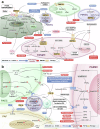
BMPR2 and TGF-β signaling pathways in PH. a BMPR2 signaling pathway and targeted therapy in PH. Cathepsin L, hypoxia, MCT, and direct BMPR2 knockdown or mutations, inhibit BMPR2 signaling, which regulates GSDME, P53/PGC1-α, NOTCH, ERK, JNK, p38, PPARγ, miRNAs, STAT3, and SOD/ROS, leading to apoptosis or pyroptosis of PAECs and PMVECs in the early stage of PH, followed by hyperproliferation of PAECs, PMVECs and PASMCs. Additionally, EndMT, ECM remodeling (COL4 and COMP), contractile phenotype inhibition of PASMCs, and inflammation (IL-6, TNF, and HMGB1) are involved in PH progression. FK506, enzastaurin, follistatin, elafin, cathepsin l shRNA, seralutinib, AAV1.BMPR2, AAV1.SIN3a, isorhamnetin, GP130, DHEA, HJC0152, BMP9, RhBMP9 alleviate PH by upregulating or activating BMPR2 and its related pathways. b TGF-β signaling pathway and targeted therapy in PH. Su/Hx, galectin-3, Hypoxia, EVs derived from HIV-infected macrophages, IL-1β, H₂O₂, MCT and NBL1 upregulate TGF-β, which targets Smad2/3/4, or p38, decrease COL1, FN1, α-SMA, vimentin, OPN and PCNA, and increase Bcl-2, leading to ECM remodeling, proliferation, migration, anti-apoptotic, EndMT. Sotatercept, ginsenoside Rg1, aspirin, danshensu, berberine, IPA, pioglitazone, IN-1233, SB525334, and SD-208 mitigate PH by modulating TGF-β signaling. PASMC pulmonary artery smooth muscle cell, PAEC pulmonary artery endothelial cell, PMVEC pulmonary microvascular endothelial cell, PAF pulmonary artery fibroblast, BMPR2 bone morphogenetic protein receptor type 2, TGF-β transforming growth factor-β, FHIT fragile histidine triad, INHBA inhibin-β-A, ActA activin-A, GSDME gasdermin E, PGC-1α peroxisome proliferator-activated receptor gamma coactivator1-alpha, TFAM transcription factor A mitochondrial, Δψm mitochondrial membrane potential, ILK integrin-linked kinase, Smad small mothers against decapentaplegic, PFKFB3 6-phosphofructo-2-kinase/fructose-2,6-biphosphatase 3, ID1 inhibitor of differentiation 1, CRYAB a-crystallin B, nmMLCK non-muscle myosin light chain kinase, HMGA1 high mobility group at-hook 1, ERK extracellular signal-regulated kinase, JNK c-Jun N terminal kinase, MCT monocrotaline, NF-κB nuclear factor kappa B, CircGSAP circular RNA-γ-secretase-activating protein, PPARγ peroxisome proliferator-activated receptorγ, PFKP phosphofructokinase 1 platelet isoform, Drp1 dynamin-related protein 1, HMGB1 high-mobility group box 1, TLR4 Toll-like receptor-4, COMP cartilage oligomeric matrix protein, SIN3a switch-independent 3a, DHEA dehydroepiandrosterone, HJC0152 a STAT3 inhibitor, GP130 Glycoprotein 130, Su/Hx SU5416-hypoxia, GDF growth and differentiation factor, FN1 fibronectin1, OPN osteopontin, COL1 type I collagen, ET-1 endothelin-1, PCNA proliferating cell nuclear antigen, NBL1 neuroblastoma suppressor of tumorigenicity 1, EV extracellular vesicle, TNF tumor necrosis factor, LTB4 leukotrieneB4, IPA Inactivated Pseudomonas aeruginosa, 5-HT 5-hydroxytryptamine, 5-LO 5-lipoxygenase, EndMT endothelial-to-mesenchymaltransition, Caspase cysteinyl aspartate specific proteinase, miR microRNA, COL4 collagen type IV
Fig. 3
Fig. 3
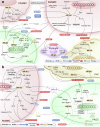
HIF signaling pathway and targeted therapy in PH. a The involvement of the HIF signaling pathway in cell proliferation, apoptosis, anti-apoptosis, ECM remodeling, migration, and cell-cell interactions in PH. Hypoxia and Tie2Cre-mediated Egln1 deletion upregulate HIF-1α and HIF-2α, leading to increased ECM protein deposition. In PASMCs, this results in the upregulation of PCNA and intracellular calcium ion concentration, along with the downregulation of Caspase-3/7/9, promoting cell proliferation, migration, and resistance to apoptosis. In PAECs, the same conditions induce the upregulation of Caspase-3, promoting apoptosis. Luteolin, PT2567, PT2385 and anti-CD146 antibody mitigate PH by regulating HIF signaling. b HIF signaling pathway also plays a crucial role in EndMT, glycolysis, inflammation, contraction, and cell-cell interactions in PH. Hypoxia, Su/Hx, and cobalt chloride upregulate HIF-1α and HIF-2α, resulting in metabolic reprogramming (e.g., PDHK), upregulation of IL and COL1, promoting inflammation, EndMT, which influences anti-apoptosis, proliferation, and migration. Conversely, the downregulation of HIF-1α in IPAH patients may lead to PASMC contraction. 2-Methoxyestradiol and apigenin mitigate PH by regulating HIF signaling. PAEC pulmonary artery endothelial cell, PASMC pulmonary artery smooth muscle cell, PAF pulmonary artery fibroblast, HIF-1α hypoxia-inducible factor-1 alpha, HIF-2α hypoxia-inducible factor-2 alpha, COL1 type I collagen, KLF5 Kruppel-like factor 5, Circ-myh8 circ_chr11_67292179-67294612, NF-κB NF-kappaB, PPARγ peroxisome proliferator-activated receptor gamma, STAT3 signal transducer and activator of transcription 3, E2F3 E2F transcription factor 3, HO-1 heme oxygenase-1, Bcl-2 B-cell lymphoma 2, Bax Bcl-2 associated X, ATG7 autophagy-related gene 7, VEGFR-2 vascular endothelial growth factor receptor 2, VEGFA vascular endothelial growth factor A, PCNA proliferating cell nuclear antigen, Arg-2 arginase-2, TSP1 thrombospondin 1, IL-33/ST2 interleukin 33/the suppression of tumorigenicity 2 receptor, NCOA6 nuclear receptor co-activator 6, PHB2 prohibitin 2, RRP1B ribosomal RNA processing 1 homolog B, AREG amphiregulin, EGFR epidermal growth factor receptor, BAD BCL2-associated agonist of cell death, FA focal adhesion, IL-6 interleukin 6, IL-1β interleukin-1beta, Twist1 Twist-related protein 1, ICAM-1 intercellular adhesion molecule-1, CXCR4 C-X-C chemokine receptor 4, SDF-1 stromal cell-derived factor 1, EDN1 endothelin 1, APLNR apelin receptor, NAMPT nicotinamide phosphoribosyltransferase, PECAM1 platelet endothelial adhesion molecule 1, ROS reactive oxygen species, NOX1 NADPH oxidase 1, PKC-α protein kinase C alpha, RASSF1 Ras association domain family 1, Drp1 dynamin-related protein 1, Kv1.5 Kv1.5 channels, PDHK pyruvate dehydrogenase kinase, IPAH diopathic pulmonary arterial hypertension, MLC myosin light chain, ET-1 endothelin-1, SM22 smooth muscle 22, PHD proline hydroxylase, Smad3 small mother against decapentaplegic family member 3, CXCL12 C-X-C chemokine ligand 12, α-SMA α-smooth muscle actin, CCL2 C-C motif ligand 2, CoCl2 cobalt chloride, Su/Hx SU5416-hypoxia, Egln1Tie2 Tie2Cre-mediated Egln1 deletion, PDGF platelet-derived growth factor, Snail Snail family transcriptional repressor, AK4 adenylate kinase 4, VEGF vascular endothelial growth factor, [Ca2+]i intracellular Ca2+ concentrations, miR microRNA, p65 a protein subunit of NF-kappaB, Caspase cysteinyl aspartate specific proteinase, siRNA small interfering RNA, CD146 cluster of differentiation 146, Ca2+ calcium ion, p53 tumor protein p53
Fig. 4
Fig. 4
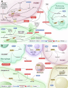
MAPK and PI3K/Akt signaling pathways in PH. a MAPK signaling pathway and targeted therapy in PH. MAPK pathway is activated by cytokines and other stimuli, leading to the upregulation of cell cycle proteins and subsequently promoting proliferation. The ERK pathway inhibits cell apoptosis, while the p38 pathway promotes cell apoptosis. Activated p38 signaling contributes to mitochondrial dysfunction and also affects inflammation by inducing the secretion of inflammatory factors. ASK1 activates p38 and JNK, thereby stimulating PAF activation, migration, and proliferation. Paeoniflorin, and 3PO alleviate PH by inhibiting ERK and its related pathways, paeoniflorin by inhibiting p38 and its related pathways, and GS-444217 alleviate PH by inhibiting ASK1/JNK/p38 axis. PASMC pulmonary artery smooth muscle cell, PAEC pulmonary artery endothelial cell, PAF pulmonary artery fibroblast, Δψm mitochondrial membrane potential, ERK extracellular signal-regulated kinase, JNK c-Jun N terminal kinase, PPARγ peroxisome proliferator-activated receptor γ, PFKFB3 6-phosphofructo-2-kinase/fructose-2,6-biphosphatase 3, PDGF platelet-derived growth factor, ET-1 endothelin-1, PCNA proliferating cell nuclear antigen, 5-HT 5-hydroxytryptamine, EETs epoxyeicosatrienoic acids, Ano1 Anoctamin-1, NPR-C atrial natriuretic peptide clearance receptor, VEGF vascular endothelial growth factor, FGF2 fibroblast growth factor 2, ASK1 apoptosis signal-regulating kinase 1, ROS reactive oxygen species, MK2 mitogen-activated protein kinase -activated protein kinase 2, Elk-1 ETS-like transcription factor, IL interleukin, Egr-1 early growth response protein 1, Bcl-2 B-cell lymphoma 2, Bax Bcl-2 associated X, MSK1 mitogen and stress-activated kinase 1, DUSP1 dual specificity phosphatase-1, SphK1 sphingosine kinase 1, Caspase cysteinyl aspartate specific proteinase, miR microRNA, 3PO 3-(4-(trifluoromethyl phenyl)-1H-pyrazole. b PI3K/Akt signaling pathway and targeted therapy in PH. CTRP9 downregulation reduces PI3K/Akt in PAECs, causing apoptosis, inflammation (ET-1, MMP-2), and dysfunction. In late PH, miR-371b-5p/PTEN downregulation activates PI3K/Akt, promoting proliferation and oxidative stress (eNOS/NO) in PAECs. BMP4-induced activation of BMPR2/PI3K/Akt in PASMCs activates PI3K/Akt in PASMCs. PI3K/Akt activation in PH drives inflammation (NF-κB) and proliferation (Smad1/5/8, FOXO3a, Cyclin A) in PAECs and PASMCs. Inhibitors like Nobiletin and Resveratrol mitigate PH, while agent like Genistein counteract early PH. PI3K phosphoinositide 3-kinase, Akt protein kinase B, CTRP9 C1q/TNF-related protein 9, eNOS endothelial nitric oxide synthase, MMP-2 matrix metalloproteinase-2, BMP4 bone morphogenetic protein 4, BMPR2 bone morphogenetic protein receptor type 2, PTEN phosphatase and tensin homolog, NF-κB nuclear factor kappa B, ECM extracellular matrix, MCT monocrotaline, IPAH idiopathic pulmonary arterial hypertension, CircDiaph3 circular RNA diaphanous-related formin 3, IGFIR insulin-like growth factor 1 receptor, miR microRNA
Fig. 5
Fig. 5
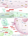
NF-κB and NLRP3 signaling pathways in PH. a NF-κB signaling pathway and targeted therapy in PH. PDGF, hypoxia, MCT and cigarette smoke activate NF-κB in PASMCs and PAECs, promoting proliferation, resistance to apoptosis, and inflammation via p65 nuclear translocation, HIF-1α activation, and CaSR upregulation. In hypoxia-PH, NLRC3 and PGC-1α are reduced, leading to activation of p65 in PAECs, which drives apoptosis, migration, and EndMT. In IPAH, NF-κB is activated in macrophages, promoting inflammation. NF-κB inhibition can alleviate PH using agents like IMD-0354, Simvastatin, Atorvastatin, Ruscogenin, Prednisolone, ad-A20, BAY11-7082, Nicorandil, and TRE. IKK IκB kinase, IκB inhibitor of NF-κB, HIF-1α hypoxia-inducible factor 1-alpha, EV extracellular vesicle, HDAC10 histone deacetylase 10, CaSR calcium-sensing receptor, NLRC3 NLR family CARD domain-containing protein 3, PGC-1α peroxisome proliferator-activated receptor gamma coactivator-1 alpha, EndMT endothelial-to-mesenchymal transition, IPAH idiopathic pulmonary arterial hypertension, NF-κB nuclear factor kappa B, PASMC pulmonary artery smooth muscle cell, PAEC pulmonary artery endothelial cell, TRE triterpenoid, TGF-β transforming growth factor-β, VPO1 vascular peroxidase 1, PTPL1 protein tyrosine phosphatase L1, HOCl hypochlorous acid, MCT monocrotaline. b NLRP3 signaling pathway and targeted therapy in PH. Su/Hx-PAH activates NLRP3 in PAECs, causing proliferation, apoptosis resistance, and pyroptosis. In macrophages, this trigger activates NLRP3, promoting inflammation and IL-1β release. In IPAH and Su/Hx-PAH, NLRP3 is activated in PASMCs, inducing pyroptosis and proliferation. In MCT-PH, NLRP3 activation in monocytes and cardiomyocytes drives hypertrophy and mitochondrial dysfunction. NLRP3 activation contributes to PH progression. PNU-282987 and Astragaloside IV inhibit NLRP3 in macrophages and PAECs. PAEC pulmonary artery endothelial cell, ROS reactive oxygen species, NLRP3 NLR family pyrin domain containing 3, Su/Hx-PAH SU5416-hypoxia-induced pulmonary arterial hypertension, GPR146 G-protein coupled receptor 146, MCT monocrotaline, HMGB1 high-mobility group box 1, STING stimulator of interferon genes, IL-1β interleukin-1 beta, SOD2 superoxide dismutase 2, IPAH idiopathic pulmonary arterial hypertension, MCT-PH monocrotaline-induced pulmonary hypertension, PNU-282987 a selective α7-nicotinic acetylcholine receptor agonist, Astragaloside IV an active compound from Astragalus membranaceus
Fig. 6
Fig. 6
Notch and AMPK signaling pathways in PH. a Notch signaling pathway and targeted therapy in PH. BMPR2 mutations in PASMCs increase TNF, reducing BMPR2 and activating Notch2. Hypoxia induces the expression of lncRNA Tug1, which activates Notch1 signaling, thereby promoting the migration of PASMCs. Notch3 activation leads PASMC migration. Additionally, it can cause the release of NICD3, which downregulates p27kip1, further enhancing PASMC proliferation. In PAECs, DLL4 nAbs impair barrier function, and hypoxia activates Notch1 to promote proliferation. Notch signaling contributes to PH, with anti-TNF and Propylthiouracil as potential therapies. PASMC pulmonary artery smooth muscle cell, BMPR2 bone morphogenetic protein receptor type 2, TNF tumor necrosis factor, miR microRNA, PAH pulmonary arterial hypertension, Notch1/2/3 Notch receptors 1/2/3, FOXC1 forkhead box C1, NICD Notch intracellular domain, SKP2 S-phase kinase-associated protein 2, Hes1 hairy and enhancer of split 1, p27kip1 cyclin-dependent kinase inhibitor 1B, PAEC pulmonary artery endothelial cell, DLL4 Delta-like ligand 4, N1-ICD Notch1 intracellular domain, p21 cyclin-dependent kinase inhibitor 1A, Bcl-2 B-cell lymphoma 2, survivin Baculoviral IAP repeat-containing 5, HPAH heritable pulmonary arterial hypertension, lncRNA long non-coding RNA, Tug1 taurine-upregulated gene 1. b AMPK signaling pathway and targeted therapy in PH. In hypoxia-PH, miR-663b is upregulated in macrophages, releasing exosomes that suppress AMPK in PASMCs, driving inflammation, oxidative stress, and proliferation. In IPAH PASMCs, NOX4 activates mTOR, inhibiting AMPK and promoting proliferation. In PAECs, suppressed AMPK in PPHN impairs angiogenesis and mitochondrial function. Metformin upregulate AMPK, mitigating PH progression. AMPK AMP-activated protein kinase, PH pulmonary hypertension, PASMC pulmonary artery smooth muscle cell, PAEC pulmonary artery endothelial cell, miR microRNA, IPAH idiopathic pulmonary arterial hypertension, NOX4 NADPH oxidase 4, mTOR mechanistic target of rapamycin, SIRT1 sirtuin 1, YAP Yes-associated protein, FOXM1 forkhead box M1, Cyclin D cyclin D1, Gal-3 galectin-3, PGC-1α peroxisome proliferator-activated receptor gamma coactivator 1-alpha, ETC electron transport chain, PPHN persistent pulmonary hypertension of the newborn, ATP adenosine triphosphate, PAH pulmonary arterial hypertension
Fig. 7
Fig. 7
Wnt signaling pathway and targeted therapy in PH. In hypoxia-PH, hyperoxia and IPAH, the Wnt/β-catenin pathway is activated, binding to the transcription factor TCF4 and leading to pro-proliferative and anti-apoptotic effects. Activated Wnt/β-catenin also promotes EndMT and ECM remodeling. Activation of the Wnt/β-catenin pathway in PAF ultimately promotes ECM remodeling. Additionally, in PAH, Wnt/PCP signaling is inhibited in PAECs and pericytes, suppressing EndMT. Naked cuticle homolog 1 and ponatinib mitigate PH by inhibiting Wnt/β-catenin pathway. PASMC pulmonary artery smooth muscle cell, PAEC pulmonary artery endothelial cell, PAF pulmonary artery fibroblast, BMPR2 bone morphogenetic protein receptor type 2, PCP planar cell polarity, EndMT endothelial-to-mesenchymaltransition, α-SMA α-smooth muscle actin, IPAH idiopathic pulmonary arterial hypertension, ECM extracellular matrix, TCF4 transcription factor 4, ChemR23 chemerin chemokine-like receptor 1, RvE1 Resolvin E1, CTEPH chronic thromboembolic pulmonary hypertension, SM22 smooth muscle protein 22, PH-LHD left ventricular secondary pulmonary hypertension, Su/Hx SU5416-hypoxia, CTGF connective tissue growth factor, WISP-1 Wnt-induced signaling protein 1, PCNA proliferating cell nuclear antigen, Egr-2 early growth response 2, LRP5/6 low-density lipoprotein-related receptors 5 and 6, FN1 fibronectin1, ERK extracellular signal-regulated kinase, ROR2 receptor tyrosine kinase-like orphan receptor type 2, COL1 type I collagen, Drp1 dynamin-related protein 1, FABP5 fatty acid-binding protein 5, OPA optic atrophy 1, OPN osteopontin
Fig. 8
Fig. 8
BMPR2 and TGF-β crosstalk with other signaling pathways in PH. a, b The networks of BMPR2 and other signaling pathways in PH. In PAECs, reduced BMPR2 signaling activates TGF-β, triggering EndMT and inflammation through Smad1/5, Smad2/3, and p38 pathways. Additionally, reduced BMPR2 signaling activates ERK1/2 pathways, promoting anti-apoptosis. In PASMCs, reduced BMPR2 signaling, influenced by ET-1 and HMGB1-induced apoptosis, supports proliferation and survival via the TGF-β/Smad1/5/8, and p38 pathways. Hypoxia and MCT stimulate HIF-1α and NF-κB, reducing BMPR2 expression through miR-130a, which promotes EndMT, inflammation, and apoptosis by suppressing β-catenin pathways in PAECs. In PASMCs, PH-induced TNF downregulates BMPR2 signaling, driving cell proliferation and glycolysis through the activation of BMP6/ALK2 and β-catenin, while suppressing miR-124-3p. c The networks of TGF-β and other signaling pathways in PH. Factors such as TGF-β, PDGF, hypoxia, and the condition of COPD contribute to cell proliferation, inflammation, EndMT, ECM remodeling, fibrosis, and myofibroblast differentiation across various cell types (PAECs, PASMCs, PAFs and RVFs), primarily through the activation of the TGFβR and its interactions with key pathways like PI3K/Akt, MAPKs, HIF-1α, and Wnt/β-catenin. PASMC pulmonary artery smooth muscle cell, BMPR2 bone morphogenetic protein receptor type 2, TGF-β transforming growth factor beta, TGFβR transforming growth factor beta receptor, PAEC pulmonary artery endothelial cell, ET-1 endothelin-1, HMGB1 high-mobility group box 1, Akt protein kinase B, ERK1/2 extracellular signal-regulated kinase 1/2, p38 mitogen-activated protein kinase, NF-κB nuclear factor kappa-light-chain-enhancer of activated B cells, HIF-1α hypoxia-inducible factor 1-alpha, MAPKs mitogen-activated protein kinases, miR microRNA, ID inhibitor of differentiation, PI3K phosphoinositide 3-kinase, Notch3 Notch receptor 3, PDGF platelet-derived growth factor, ROS reactive oxygen species, Wnt5a/b Wnt family member 5A/B, COPD chronic obstructive pulmonary disease, IPAH idiopathic pulmonary arterial hypertension, ECM extracellular matrix, EndMT endothelial-to-mesenchymal transition, Skp2 S-phase kinase-associated protein 2, Hes1 hairy and enhancer of split 1, p27Kip1 cyclin-dependent kinase inhibitor 1B, N1-ICD Notch1 intracellular domain, p21 cyclin-dependent kinase inhibitor 1A, Bcl-2 B-cell lymphoma 2, survivin Baculoviral IAP repeat-containing 5
Fig. 9
Fig. 9
PI3K, NF-κB and AMPK crosstalk with other signaling pathways in PH. a The networks of PI3K and other signaling pathways in PH. In PAECs and PASMCs, IPAH, hypoxia, and 5-HT activate ERK, PI3K, JNK, and calcium signaling, promoting proliferation and inflammation through the Akt pathway. b The networks of NF-κB and other signaling pathways in PH. Hypoxia and FGF2 activate NF-κB p65 signaling in PAECs and PASMCs through interactions with key pathways, including HIF-1α, p38, and ERK. These networks drive processes such as inflammation, proliferation, angiogenesis, and EndMT. In RVF, MCT induces right ventricular failure through NF-κB activation and its interactions with MAPK signaling pathways. c The networks of AMPK and other signaling pathways in PH. In PAECs and PASMCs, hypoxia and PPHN suppress AMPK signaling, leading to the activation of key pathways such as Notch, NF-κB, NLRP3, and Akt, which drive angiogenesis, autophagy, inflammation, pyroptosis, and increased cell proliferation. IPAH idiopathic pulmonary arterial hypertension, PAEC pulmonary artery endothelial cell, PASMC pulmonary artery smooth muscle cell, 5-HT serotonin, ERK extracellular signal-regulated kinase, PI3K phosphoinositide 3-kinase, JNK c-Jun N-terminal kinase, Akt protein kinase B, FGF2 fibroblast growth factor 2, HIF-1α hypoxia-inducible factor 1-alpha, MAPK mitogen-activated protein kinase, RVF right ventricular failure, NF-κB nuclear factor kappa-light-chain-enhancer of activated B cells, MCT monocrotaline, PPHN persistent pulmonary hypertension of the newborn, AMPK AMP-activated protein kinase, EndMT endothelial-to-mesenchymal transition

References

    1. Ruopp, N. F. & Cockrill, B. A. Diagnosis and treatment of pulmonary arterial hypertension: a review. JAMA327, 1379–1391 (2022). - PubMed
    1. Humbert, M. et al. 2022 ESC/ERS Guidelines for the diagnosis and treatment of pulmonary hypertension. Eur. Heart J.43, 3618–3731 (2022). - PubMed
    1. Cahal, D. A. Appetite suppressants and pulmonary hypertension. Lancet1, 947 (1969). - PubMed
    1. Mocumbi, A. et al. Pulmonary hypertension. Nat. Rev. Dis. Prim.10, 1 (2024). - PubMed
    1. Rabinovitch, M., Guignabert, C., Humbert, M. & Nicolls, M. R. Inflammation and immunity in the pathogenesis of pulmonary arterial hypertension. Circ. Res.115, 165–175 (2014). - PMC - PubMed

MeSH terms

LinkOut - more resources