. 2025 Jun 30;16(1):5376.
doi: 10.1038/s41467-025-61134-0.

Monkeypox virus spreads from cell-to-cell and leads to neuronal death in human neural organoids

Affiliations

Monkeypox virus spreads from cell-to-cell and leads to neuronal death in human neural organoids

Isabel Schultz-Pernice et al. Nat Commun. .

Abstract

In 2022-23, the world witnessed the largest recorded outbreak of monkeypox virus (MPXV). Neurological manifestations were reported alongside the detection of MPXV DNA and MPXV-specific antibodies in the cerebrospinal fluid of patients. Here, we analyze the susceptibility of neural tissue to MPXV using human neural organoids (hNOs) exposed to a clade IIb isolate. We report susceptibility of several cell types to the virus, including neural progenitor cells and neurons. The virus efficiently replicates in hNOs, as indicated by the exponential increase of infectious viral titers and establishment of viral factories. Our findings reveal focal enrichment of viral antigen alongside accumulation of cell-associated infectious virus, suggesting viral cell-to-cell spread. Using an mNeonGreen-expressing recombinant MPXV, we confirm cell-associated virus transmission. We furthermore show the formation of beads in infected neurites, a phenomenon associated with neurodegenerative disorders. Bead appearance precedes neurite-initiated cell death, as confirmed through live-cell imaging. Accordingly, hNO-transcriptome analysis reveals alterations in cellular homeostasis and upregulation of neurodegeneration-associated transcripts, despite scarcity of inflammatory and antiviral responses. Notably, tecovirimat treatment of MPXV-infected hNOs significantly reduces infectious virus loads. Our findings suggest that viral disruption of neuritic transport drives neuronal degeneration, potentially contributing to MPXV neuropathology and revealing targets for therapeutic intervention.


Conflict of interest statement

Competing interests: The authors declare no competing interests.

Figures

Fig. 1
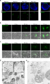
Fig. 1. MPXV has a broad cellular tropism in hNOs and 2D neural cultures.
a Representative micrographs showing the cell diversity and morphology of ESC-derived hNOs at selected developmental time points (10, 60, and 112 days). Selected markers of NPCs (SOX2, pink) and neurons (TUJ1, orange) are presented. DAPI, blue. Scale bar, 50 µm. b Representative micrographs illustrating the target cells of MPXV in 70-75 days old hNOs 10 days p.i. with a clade IIb MPXV isolate at an MOI of 0.1 TCID50/cell. DAPI, blue; MPXV, green; SOX2, pink; TUJ1, orange. Scale bar, 10 µm. Characterization of 2D NPC (c) and forebrain neuron (d) cultures. DAPI, blue; SOX2, pink; TUJ1, orange. Scale bar, 100 μm. Exemplary image of 2D-cultured NPCs (e) and neurons (f) 8 h p.i. with a clade IIb MPXV isolate at an MOI of 0.01 TCID50/cell. DAPI, blue; MPXV, green; SOX2, pink; TUJ1, orange. Scale bar, 10 μm.
Fig. 2
Fig. 2. Productive MPXV infection in hNOs and 2D cultured neural cells.
a Representative micrographs of hNOs infected with a clade IIb MPXV isolate at a rate of 0.1 TCID50/cell at 83 days of development. Infection was followed by immunofluorescence from 2 to 14 days p.i. by imaging of sectioned hNOs. DAPI, blue; MPXV, green. Scale bar, 1000 µm. b Representative micrographs depicting infection progression of 2D cultured NPCs challenged with an mNG-expressing clade IIb MPXV reporter construct at a rate of 0.01 TCID50/cell. Scale bar, 100 μm. c Representative images of 2D cultured forebrain neurons exposed to an mNG-expressing clade IIb MPXV construct at a rate of 0.01 TCID50/cell. Scale bar, 100 μm. d, e Representative images acquired by transmission electron microscopy at 10 days p.i. of 95 days old hNOs infected with a clade IIb MPXV isolate with an MOI of 0.1 TCID50/cell. d’ Magnification demonstrating the presence of intracellular mature virions within cells. Scale bar, 1 µm. d” Magnification showing the presence of viral particles at different stages of development alongside perinuclear viral factories, displaying cytoskeletal condensations. Scale bar, 1 µm. e Illustration of degenerative signs, bursting and release of intracellular compartment containing high amounts of monkeypox virions. Scale bar, 1 µm.
Fig. 3
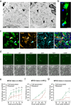
Fig. 3. MPXV spreads through cell-to-cell contact.
a Representative transmission electron microscopy images of 95 days old hNOs at 10 days p.i. exposed to a clade IIb MPXV isolate at an MOI of 0.1 TCID50/cell. MPXV particles were observed localizing to neurites in infected tissue. Black arrowheads indicate exemplary virion-harboring neurites. Scale bar, 0.5 µm. b Representative image of detection by immunofluorescence of MPXV signal within neural filaments of interconnected, distinct cells. DAPI, blue; MPXV, green. Scale bar, 10 µm. c Representative micrographs of 95-day-old hNOs infected with a clade IIb MPXV isolate, showing the filaments connecting MPXV antigen-positive cells and displaying diverse TUJ1 and F-actin patterns. Some filaments were observed showing expression of both markers (yellow arrowheads), indicative of their neuritic nature, others lacked TUJ1 signal, indicative of TNTs (blue arrowheads). DAPI, blue; MPXV, green; TUJ1, orange; F-actin, cyan. Scale bar, 10 µm. d Representative micrographs of 2D cultured forebrain neurons exposed to an mNG-expressing clade IIb MPXV reporter construct, showing viral dissemination between interconnected neighboring cells. White arrowheads indicate mNG-harboring neurites. Images were taken at 40-min intervals, time represented in minutes. Scale bar, 100 µm. e Cell-associated and released infectious MPXV 2 to 14 days p.i. of 70–95 days old hNOs infected with a clade IIb isolate at a rate of 0.1 TCID50/cell. Each symbol represents an individual organoid (n = 7–8 independent organoid batches). Unpaired, two-tailed student’s t-test was applied to compare groups. M indicates mock. f Cell-associated and released MPXV titers in 2D cultured NPCs challenged with a clade IIb MPXV isolate at a rate of 0.01 TCID50/cell. Each symbol represents one NPC batch (n = 3 independent NPC batches). Unpaired, two-tailed student’s t-test was applied to compare groups. M indicates mock. g Released and cell-associated infectious MPXV in 2D cultured forebrain neurons exposed to a clade IIb MPXV isolate at a rate of 0.01 TCID50/cell. Each symbol represents one neuron batch (n = 4 independent neuron batches). Unpaired, two-tailed student’s t-test was applied to compare groups. M indicates mock. Dashed line indicates the detection limit of the assay. Source data are provided as a Source Data file.
Fig. 4
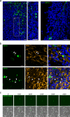
Fig. 4. MPXV causes neuritic beading and neuron death.
a, b Immunofluorescence analysis 8-14 days p.i. of 70 days old hNOs exposed to a clade IIb MPXV isolate at a rate of 0.1 TCID50/cell. a Representative micrographs showing viral antigen accumulating within regularly interspaced swellings (beads) in filaments, spanning over long distances within the tissue. DAPI, blue; MPXV, green. Scale bar, 25 µm. b Representative micrographs of beaded filaments harboring monkeypox virions co-localizing with the neuronal marker TUJ1 in both filaments and beads. White arrowheads indicate regions displaying marker co-localization. DAPI, blue; MPXV, green; TUJ1, orange. Scale bar, 10 µm. c Representative micrographs of 2D forebrain neurons exposed to an mNG-expressing clade IIb MPXV reporter construct displaying neuritic beading in filaments of a virus-harboring cell in concomitance with sudden mNG signal loss in the cell’s soma. mNG signal is subsequently observed to progressively diminish while beads still harbor detectable reporter protein signal. Images were taken at 30-min intervals, time represented in minutes. Scale bar, 100 µm.
Fig. 5
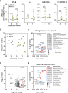
Fig. 5. Transcriptional profile alteration in hNOs during MPXV infection.
a Fold change of transcriptional expression levels of selected proinflammatory cytokine and chemokine genes including IFN-β, IL-6, IL-8/CXCL8, and IP-10/CXCL10 in 74-93 days old hNOs exposed to a MPXV clade IIb isolate at a rate of 0.1 TCID50/cell. Boxplots indicate the median value (centerline) and interquartile ranges (box edges), with whiskers extending to the lowest and the highest values. Each symbol represents an individual organoid (n = 3 independent organoid batches). Two-tailed Mann-Whitney U test was applied to compare groups. be Transcriptomic analysis of MPXV-challenged versus mock-treated hNOs. Organoids were infected with a MPXV clade IIb isolate at a rate of 0.1 TCID50/cell at 74–93 days of development (n = 3 independent organoid batches). hNOs were collected for RNA isolation at 4 days p.i. b PCA showing separate clustering of mock and MPXV-exposed organoids. c Volcano plot representing DEGs between MPXV-challenged and mock-treated hNOs. Two-tailed Wald test corrected for multiple comparisons using a Benjamini & Hochberg correction was applied; padj < 0.05. d GO analysis results displaying top 7 up- and downregulated biological processes in MPXV-infected versus mock-treated hNOs. One-tailed Fisher’s exact test corrected for multiple comparisons using a Benjamini Hochberg was applied; padj < 0.05. e GO analysis results displaying top 7 up- and downregulated molecular functions in MPXV-exposed and mock-treated hNOs. One-tailed Fisher’s exact test corrected for multiple comparisons using a Benjamini Hochberg was applied; padj < 0.05. Source data are provided as a Source Data file.
Fig. 6
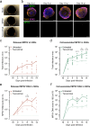
Fig. 6. Tecovirimat treatment limits MPXV spread in hNOs.
a Representative micrographs of sectioned mock-treated (top) and MPXV-exposed hNOs cultured in regular (center) or tecovirimat-complemented medium (bottom) at 14 days p.i. Organoids were exposed to a clade IIb MPXV isolate at a rate of 0.1 TCID50/cell at 84 days of development. Tecovirimat was added to the medium 2 h after initial MPXV-exposure, to a final concentration of 1 µM. Scale bar, 2000 μm. b Representative sections of tecovirimat-treated hNOs sampled at selected time points p.i. showing unaltered tissue organization and limited virus antigen presence. Infected organoids were exposed to a clade IIb MPXV isolate at a rate of 0.1 TCID50/cell at 84 days of development. DAPI, blue; MPXV, green; TUJ1, orange; SOX2, pink. Scale bar, 1000 μm. Released (c) and cell-associated (d) infectious MPXV 2 to 14 days p.i. of 70-95 days old hNOs infected with a clade IIb isolate at a rate of 0.1 TCID50/cell and cultured in regular or tecovirimat-supplemented medium. Each symbol represents an individual organoid (n = 7-8 independent organoid batches). Unpaired, two-tailed student’s t-test was applied to compare groups. M indicates mock. Released (e) and cell-associated (f) MPXV loads detected through qPCR analysis in samples taken 2 to 14 days p.i. 84-85 days old hNOs were exposed to a clade IIb MPXV isolate at a rate of 0.1 TCID50/cell and cultured in regular or tecovirimat-supplemented medium. Each symbol represents an individual organoid (n = 3 independent organoid batches). Unpaired, two-tailed student’s t-test was applied to compare groups. M indicates mock. Dashed line indicates the detection limit of the assay(s). Source data are provided as a Source Data file.

References

    1. World Health Organization. 2022 Mpox (Monkeypox) Outbreak: Global Trends. Vol. 2023 (World Health Organization, 2022).
    1. World Health Organization. Monkeypox - United Kingdom of Great Britain and Northern Ireland. Vol. 2023 (World Health Organization, 2022).
    1. Thornhill, J. P. et al. Monkeypox Virus infection in humans across 16 countries - April-June 2022. N. Engl. J. Med.387, 679–691 (2022). - PubMed
    1. Perez Duque, M. et al. Ongoing monkeypox virus outbreak, Portugal, 29 April to 23 May 2022. Eurosurveillance27, 2200424 (2022). - PMC - PubMed
    1. Girometti, N. et al. Demographic and clinical characteristics of confirmed human monkeypox virus cases in individuals attending a sexual health centre in London, UK: an observational analysis. Lancet Infect. Dis.22, 1321–1328 (2022). - PMC - PubMed