[Preprint]. 2025 Jun 17:2025.06.11.659204.
doi: 10.1101/2025.06.11.659204.

Selective inhibition of MR1-restricted T cell activation by a novel MR1-targeting nanobody

Affiliations

Selective inhibition of MR1-restricted T cell activation by a novel MR1-targeting nanobody

Timothy Bates et al. bioRxiv. .

Abstract

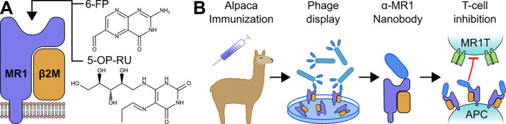
MR1 is a non-polymorphic, ubiquitously expressed, MHC class I-like antigen-presenting molecule that presents small-molecule metabolites to T cells. Studies have shown that MR1 plays a role in microbial infection, inflammation, and tumor immunity. The antigens it presents include metabolites of microbial and self-origin as well as small-molecule drugs and form stable complexes with MR1 that are displayed on the cell surface to activate T cells. However, unlike classical MHC I and II molecules, the fundamental biology of MR1 remains poorly understood, particularly the mechanisms governing antigen loading and intracellular trafficking. This knowledge gap is largely due to the lack of molecular tools available to precisely manipulate MR1 function. In this study, we describe a high-affinity (1.6 nM K D ) anti-MR1 nanobody, MR1Nb1. We characterize the binding of this nanobody including affinity by ELISA and kinetics by BLI. Crucially, we map the binding epitope of MR1Nb1 on MR1 by HDX-MS, providing key insights into the mechanism through which it blocks MR1T cell activation. In functional assays MR1Nb1 effectively and specifically blocks MAIT cell activation by cells infected with M. tuberculosis or treated with M. smegmatis supernatant. This nanobody represents a unique and versatile tool for the field, as it can be produced inexpensively and expressed intracellularly within antigen-presenting cells. Hence, our study provides a powerful new molecular probe for dissecting the mechanistic underpinnings of MR1 biology and uncover its broader roles in immunity.

Keywords: MAIT; MR1; Nanobody; T cell; tuberculosis.


Conflict of interest statement

Conflicts of Interest J.E.B. reports personal fees from Scorpion Therapeutics, Reactive therapeutics and Olema Oncology, and research grants from Novartis. T.A.B. and F.G.T. are co-founders of AlpaCure LLC. All other authors declare no competing interests.

Figures

Figure 1:
Figure 1:. Schematic diagrams.
Membrane-bound MR1 attached to its obligate binding partner, β2M. The antigen binding groove of MR1 can bind and display a variety of small molecule antigens including 6-FP and 5-OP-RU (A). Alpacas were immunized with recombinant MR1; phage display was performed on the resulting immune library; individual candidates were produced and characterized, including T-cell inhibition assays (B).
Figure 2:
Figure 2:. MR1Nb1 and MR1Nb3 bind to MR1 loaded with either 6-FP or 5-OP-RU.
ELISA EC50 curves showing MR1Nb1 and MR1Nb3 binding to plates coated with 6-FP-loaded MR1 (A), and 5-OP-RU-loaded MR1 (B). Biolayer interferometry of MR1Nb1 on 6-FP-loaded MR1 (C), and 5-OP-RU-loaded MR1 (D); then similarly with MR1Nb3 on 6-FP-loaded MR1 (E), and 5-OP-RU-loaded MR1 (F). ELISA experiments were performed in triplicate (n=3) and BLI experiments were performed in duplicate (n=2).
Figure 3:
Figure 3:. BLI showing competitive binding of MR1Nb1 with MR1Nb3, but not mAb 26.5.
MR1 loaded with 6-FP were first bound with MR1Nb1 (A) or MR1Nb3 (B) then immediately transferred to the opposing VHH or mAb 26.5. This was repeated with MR1 loaded with 5-OP-RU for MR1Nb1 (C) and MR1Nb3 (D).
Figure 4.
Figure 4.. HDX-MS analysis of the interaction of MR1 with MR1Nb1.
HDX differences (defined as >5%, 0.4 Da, and P< 0.01 in an unpaired two-tailed t test at any time point) upon MR1 binding to MR1Nb1 mapped on MR1 structure (PDB: 4GUP) (A). HDX differences of MR1 when bound to MR1Nb1 mapped on the structure of a TRBV6–4 MAIT TCR in complex with MR1 (PDB: 4PJ7) (B). Sum of the deuteron difference of MR1 upon binding to MR1Nb1 across the entire time course (C). Peptides that met the significance criteria described in (A) are colored red. Error is shown as the sum of SDs across all time points (n=3). Deuterium incorporation in MR1 peptides showing significant changes in the presence (red) and absence (black) of MR1Nb1 (D).
Figure 5:
Figure 5:. MR1Nb1 specifically blocks MR1-mediated presentation of both exogenous ligands and antigens derived from intracellular infection with Mtb.
BEAS-2B cells were pre-treated with an irrelevant control nanobody (open circles), MR1Nb1 (filled circles), an IgG2a isotype control antibody (open squares), or the anti-MR1 antibody clone 26.5 (filled squares) at the indicated concentrations, incubated with either M. smegmatis supernatant (A) or CFP102–9 peptide (B), and then co-cultured with the MR1-restricted MAIT cell clone D416-G11 (A) or the HLA-B45-restricted classical T cell clone D466-A10 (B) overnight. BEAS-2B cells were infected with Mtb strain H37Rv at an MOI of 8 overnight before incubation with 5 μg/ml of each blocking reagent and subsequent co-culture with MAIT cell clone D416-G11 (C) or classical T cell clone D466-A10 (D). IFN-γ production was measured as spot-forming units (SFU) by ELISpot. Data are the pooled means of technical duplicates from (n=3) independent experiments and shown as mean +/− standard deviation.

References

    1. Corbett AJ et al. T-cell activation by transitory neo-antigens derived from distinct microbial pathways. Nature. 2014. May;509(7500):361–5. - PubMed
    1. Harriff MJ et al. MR1 displays the microbial metabolome driving selective MR1-restricted T cell receptor usage. Sci Immunol. 2018. Jul 13;3(25):eaao2556. - PMC - PubMed
    1. Kjer-Nielsen L et al. MR1 presents microbial vitamin B metabolites to MAIT cells. Nature. 2012. Nov 29;491(7426):717–23. - PubMed
    1. Hashimoto K et al. A Gene Outside the Human MHC Related to Classical HLA Class I Genes. Science. 1995. Aug 4;269(5224):693–5. - PubMed
    1. Napier RJ et al. The Role of Mucosal Associated Invariant T Cells in Antimicrobial Immunity. Front Immunol. 2015;6:344. - PMC - PubMed

Publication types