Gel Immersion Endoscopic Submucosal Dissection Using a Scissor-type Knife for Superficial Non-ampullary Duodenal Epithelial Tumors
- PMID: 40589618
- PMCID: PMC12208102
- DOI: 10.1002/deo2.70157
Gel Immersion Endoscopic Submucosal Dissection Using a Scissor-type Knife for Superficial Non-ampullary Duodenal Epithelial Tumors
Abstract
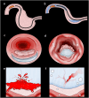
Objectives: This study aimed to compare the short-term therapeutic outcomes between conventional endoscopic submucosal dissection (C-ESD) and gel immersion ESD (GI-ESD) for superficial non-ampullary duodenal epithelial tumors (SNADETs).
Methods: A retrospective analysis was conducted on patients with SNADETs who underwent C-ESD or GI-ESD between June 2016 and May 2024. To reduce proficiency bias, the first 50 cases per endoscopist were excluded. C-ESD was performed using a scissor-type knife under CO2 insufflation, while GI-ESD was performed using the same knife under gel immersion. Primary outcomes included en bloc and R0 resection rates; secondary outcomes were resection time, adverse events, and inflammatory response.
Results: Overall, 51 C-ESD and 49 GI-ESD procedures were analyzed. Both groups achieved 100% en bloc resection. R0 resection rates were comparable (C-ESD: 92.6%, GI-ESD: 90.2%, p = 0.661). Muscle layer exposure was significantly lower in the GI-ESD group (1.9%) than in the C-ESD group (16.7%, p = 0.032). The mean white blood cell count was also significantly lower in the GI-ESD group (p = 0.038). The incidence of adverse events in the C-ESD and GI-ESD groups was 5.6% and 1.9%, respectively (p = 0.627). However, no cases of perforation or aspiration were observed in the GI-ESD group.
Conclusions: GI-ESD is a safe and effective alternative to conventional ESD for SNADETs, offering comparable resection outcomes and low risk of adverse events with a reduced risk of muscle layer exposure.
Keywords: adverse event | endoscopic submucosal dissection | gel immersion | scissor‐type knife | superficial non‐ampullary duodenal epithelial tumor.
© 2025 The Author(s). DEN Open published by John Wiley & Sons Australia, Ltd on behalf of Japan Gastroenterological Endoscopy Society.
Conflict of interest statement
Osamu Dohi, Naohisa Yoshida, and Tomohisa Takagi received research funds from Fujifilm Co., Ltd. Naohisa Yoshida received lecture fees from Fujifilm Co., Ltd. The other authors declare no conflicts of interest.
Figures


References
-
- Höchter W., Weingart J., Seib H. J., et al., “Duodenal Polyps. Incidence, Histologic Substrate and Significance,” Deutsche Medizinische Wochenschrift 109 (1984): 1183–1186. - PubMed
-
- Jepsen J. M., Persson M., Jakobsen N. O., et al., “Prospective Study of Prevalence and Endoscopic and Histopathologic Characteristics of Duodenal Polyps in Patients Submitted to Upper Endoscopy,” Scandinavian Journal of Gastroenterology 29 (1994): 483–487. - PubMed
-
- Coupland V. H., Kocher H. M., Berry D. P., et al., “Incidence and Survival for Hepatic, Pancreatic and Biliary Cancers in England Between 1998 and 2007,” Cancer Epidemiology 36 (2012): e207–14. - PubMed
-
- Yoshida M., Yabuuchi Y., Kakushima N., et al., “The Incidence of Non‐ampullary Duodenal Cancer in Japan: The First Analysis of a National Cancer Registry,” Journal of Gastroenterology and Hepatology 36 (2021): 1216–1221. - PubMed
LinkOut - more resources
Full Text Sources
Research Materials
Miscellaneous
