Review
. 2025 Jun 16:16:1595852.
doi: 10.3389/fimmu.2025.1595852. eCollection 2025.

The emerging role of cuproptosis in spinal cord injury

Affiliations
Review

The emerging role of cuproptosis in spinal cord injury

Daoran Xu et al. Front Immunol. .

Abstract

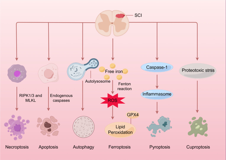
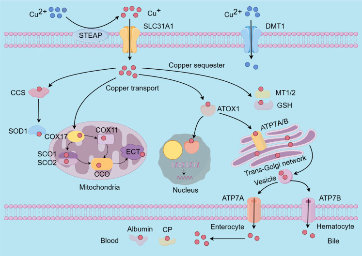
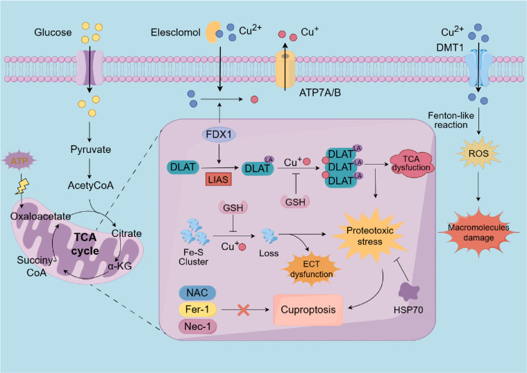
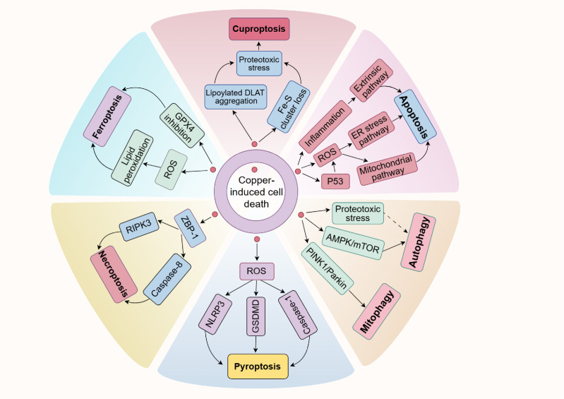
Copper is a vital trace element integral to numerous biological processes, including iron metabolism, neurotransmitter synthesis, mitochondrial respiration, oxidative stress regulation, and energy production. However, disturbances in copper metabolism can result in pathological conditions, including cuproptosis-a newly recognized form of programmed cell death (PCD) marked by copper accumulation and the disruption of copper-dependent metabolic pathways. Cuproptosis has been associated with various diseases, such as cancer, metabolic disorders and neurodegenerative disorders. In the context of spinal cord injury (SCI), multiple pathological mechanisms, including oxidative stress, inflammation, and PCD could impact the patient's prognosis with SCI. This review seeks to elucidate the pathophysiological underpinnings of SCI, the mechanisms and biological significance of copper homeostasis and the role of cuproptosis in SCI.

Keywords: copper homeostasis; cuproptosis; programmed cell death; reactive oxygen species; spinal cord injury.


Conflict of interest statement

The authors declare that the research was conducted in the absence of any commercial or financial relationships that could be construed as a potential conflict of interest.

Figures

Figure 1
Figure 1
Spinal cord injury and programmed cell death. Initially, the application of mechanical force damages the cell membrane and releases intracellular components, further triggering autophagic processes as a defense mechanism. Following this, a range of pathophysiological processes occur, including oxidative stress, inflammation, and immune response, which eventually result in the apoptosis, necroptosis, and pyroptosis of neuronal and glial cells through specific mechanisms. Moreover, an overabundance of ROS may lead to lipid peroxidation and trigger ferroptosis. It’s intriguing that an excess of copper at the injury site might induce mitochondrial proteotoxic stress and cause cuproptosis. SCI, Spinal cord injury; RIPK1/3, receptor-interacting protein kinases; MLKL, mixed lineage kinase domain-like protein; ROS, reactive oxygen species; GPX4, glutathione peroxidase 4.
Figure 2
Figure 2
The molecular mechanisms underlying copper metabolism involve several key processes. The absorption of copper ions is primarily facilitated by the transporters SLC31A1 and DMT1. Notably, SLC31A1 is specific to the uptake of monovalent copper ions, necessitating the reduction of divalent copper ions to monovalent copper ions by STEAP before uptake. Within cells, copper is transported to and interacts with specific cytoplasmic copper chaperones, including COX17, CCS, and ATOX1, which direct copper to distinct cellular compartments such as the mitochondrial electron transport chain, the trans-Golgi network, and the nucleus. Copper insertion and disulfide bond formation in SOD1 are specifically facilitated by the CCS. To mitigate the cytotoxic effects of excess intracellular copper, copper ions can bind to MT1/2 and GSH for storage. Additionally, the maintenance of intracellular copper homeostasis is achieved through the export of surplus copper ions into the bloodstream via ATP7A and ATP7B. Subsequently, copper is transported throughout the body by CP and albumin. SLC31A1, Solute carrier family 31 member 1; DMT1, divalent metal transporter 1; STEAP, six-transmembrane epithelial antigen of the prostate; COX17/11, cytochrome c oxidase copper chaperone 17/11; CCS, cytoplasmic-mitochondrial metallochaperones; ATOX1, antioxidant protein 1; MT1/2, metallothioneins; GSH, glutathione; Cu-Zn SOD1, Cu-Zn superoxide dismutase 1; SCO1/2, sythesis of cytochrome oxidase 1/2; CCO, cytochrome oxidase; ATP7A/B, ATPase 7A/B; CP, cuproenzymes.
Figure 3
Figure 3
A diagrammatic representation of the cuproptosis process. Extracellular copper is sequestered by the copper ionophore elesclomol and transported into intracellular compartments. As an upstream regulator of protein lipoylation, FDX1/LIAS may enhance the lipoylation of TCA cycle enzymes, such as DLAT. Furthermore, within the mitochondria, FDX1 facilitates the reduction of divalent copper to its monovalent state. Subsequently, the monovalent copper could interact with DLAT and Fe-S cluster proteins results in DLAT aggregation and the depletion of Fe-S cluster proteins. These aberran interactions result in the TCA cycle and ETC to malfunction, triggering cuproptosis through proteotoxic stress, which eventually disrupts ATP synthesis. Notably, HSP70 and copper chelators, such as GSH, can inhibit cuproptosis. However, NAC, ferrostatin-1, and necrostatin-1 do not mitigate copper-induced cell death. Additionally, excessive copper ions can also induce oxidative stress and elevate ROS levels, thereby damaging intracellular macromolecules. FDX1, Ferredoxin 1; LIAS, lipoic acid synthetase; TCA, tricarboxylic acid; Fe-S, iron-sulfur cluster; DLAT, dihydrolipoamide S-acetyltransferase; ETC, electron transport chain; HSP70, heat shock protein 70; GSH, glutathione; NAC, N-acetylcysteine; ferrostatin-1, ferroptosis inhibitors; necrostatin-1, necroptosis inhibitors; ROS, reactive oxygen species.
Figure 4
Figure 4
Copper is closely associated with various forms of PCD. On the one hand, an excess of copper can enhance p53 expression and ROS levels or initiate inflammation, causing cell apoptosis. On the other hand, copper exposure increases ROS levels or reduces GPX4 expression, which subsequently leads to ferroptosis or pyroptosis via specific pathways. It has been shown that copper induces necroptosis mediated by ZBP1. Furthermore, copper can also trigger autophagy and mitophagy by inhibiting the AMPK/mTOR pathway or activating the PINK1/Parkin signaling pathway, respectively. It is noteworthy that recent research has shown copper could induce cuproptosis, a newly discovered cell death type, characterized by the accumulation of lipoylated proteins and the depletion of Fe-S clusters. Possible connections are shown with dashed arrows.

Similar articles

References

    1. Chen J, Jiang Y, Shi H, Peng Y, Fan X, Li C. The molecular mechanisms of copper metabolism and its roles in human diseases. Pflugers Archiv: Eur J Physiol. (2020) 472:1415–29. doi: 10.1007/s00424-020-02412-2 - DOI - PubMed
    1. Harris ED. Copper as a cofactor and regulator of copper,Zinc superoxide dismutase. J Nutr. (1992) 122:636–40. doi: 10.1093/jn/122.suppl_3.636 - DOI - PubMed
    1. Gale J, Aizenman E. The physiological and pathophysiological roles of copper in the nervous system. Eur J Neurosci. (2024) 60:3505–43. doi: 10.1111/ejn.16370 - DOI - PMC - PubMed
    1. Vetlényi E, Rácz G. the physiological function of copper, the etiological role of copper excess and deficiency. Orvosi hetilap. (2020) 161:1488–96. doi: 10.1556/650.2020.31854 - DOI - PubMed
    1. Wang Q, Sun J, Chen T, Song S, Hou Y, Feng L, et al. Ferroptosis, pyroptosis, and cuproptosis in alzheimer’s disease. ACS Chem Neurosci. (2023) 14:3564–87. doi: 10.1021/acschemneuro.3c00343 - DOI - PubMed

LinkOut - more resources