. 2025 Jul 1;135(13):e188932.
doi: 10.1172/JCI188932.

A haploinsufficiency restoration strategy corrects neurobehavioral deficits in Nf1+/- mice

Affiliations

A haploinsufficiency restoration strategy corrects neurobehavioral deficits in Nf1+/- mice

Su Jung Park et al. J Clin Invest. .

Abstract

Neurofibromatosis type 1 (NF1) is a genetic disorder caused by mutations of the NF1 tumor suppressor gene resulting in the loss of function of neurofibromin, a GTPase-activating protein (GAP) for Ras. While the malignant manifestations of NF1 are associated with loss of heterozygosity of the residual WT allele, the nonmalignant neurodevelopmental sequelae, including autism spectrum disorder (ASD) and/or attention deficit hyperactivity disorder (ADHD) are prevalent morbidities that occur in the setting of neurofibromin haploinsufficiency. We reasoned that augmenting endogenous levels of WT neurofibromin could serve as a potential therapeutic strategy to correct the neurodevelopmental manifestations of NF1. Here, we used a combination of genetic screening and genetically engineered murine models to identify a role for the F-box protein FBXW11 as a regulator of neurofibromin degradation. Disruption of Fbxw11, through germline mutation or targeted genetic manipulation in the nucleus accumbens, increased neurofibromin levels, suppressed Ras-dependent ERK phosphorylation, and corrected social learning deficits and impulsive behaviors in male Nf1+/- mice. Our results demonstrate that preventing the degradation of neurofibromin is a feasible and effective approach to ameliorate the neurodevelopmental phenotypes in a haploinsufficient disease model.

Keywords: Genetic diseases; Genetics; Neurodevelopment; Neuroscience; Ubiquitin-proteosome system.


Figures

Figure 1
Figure 1. F-box proteins FBXW11 and FBXO3 are involved in the degradation of neurofibromin.
(A) Control (Cont.) and F-box–specific siRNAs were transfected into human diploid fibroblasts. After 72 hours, lysates were prepared and used for immunoblotting to detect NF1 (top) and pERK1/2 (bottom doublet). Red font highlights increased neurofibromin levels in lysates prepared from wells containing siRNAs targeting FBXW11 or FBXO3. An siRNA targeting FBXW1A (bTrCP1, BTRC) was not included. (B) HeLa cells were transfected with a control siRNA or with siRNAs targeting FBXO3 or FBXW11. NF1, pERK1/2, and total ERK1/2 levels were analyzed 72 hours after transfection by immunoblotting. GAPDH was used as a loading control. (C) Left: CHX was added at a final concentration of 20 μg/mL to HeLa cells for the indicated durations prior to harvesting for immunoblotting to detect NF1. GAPDH was used as a loading control. Right: NF1 levels were quantified by densitometry relative to GAPDH. *P < 0.05, by 1-way ANOVA with Dunnett’s multiple-comparison test. Data indicate the mean ± SEM. (D) Left: HeLa cells were transfected with control siRNA or siRNA targeting FBXO3 or FBXW11. Sixty-eight hours after transfection, cells were treated with CHX (20 μg/mL final concentration) for 2 hours prior to harvesting and immunoblotting to detect NF1. GAPDH was used as a loading control. Right: NF1 levels were quantified by densitometry relative to GAPDH. *P < 0.05 comparing siCont with or without CHX, by unpaired 2-tailed t test. Data indicate the mean ± SEM.
Figure 2
Figure 2. Neurofibromin levels affected by ectopic FBXW11 or FBXO3 expression or small-molecule F-box inhibitors.
(A and B) HeLa cells transfected with an empty expression vector or with vectors containing either an FBXW11 cDNA or an FBXO3 cDNA were collected and lysed 48 hours later. NF1, FBXW11, and FBXO3 proteins were detected by immunoblotting, with GAPDH used as a loading control. (C) Nf1+/– MEFs were treated with the FBXW11 inhibitor PDTC (50 μM) or the FBXO inhibitor BC-1215 (20 μg/mL) for 6 hours prior to harvesting for immunoblotting. The lower panels represent densitometric analysis comparing NF1 levels with the GAPDH control. *P < 0.05 and **P < 0.01, by unpaired 2-tailed t test. Data indicate the mean ± SEM.
Figure 3
Figure 3. FBXW11 preferentially interacts with and polyubiquitinates the GRD of neurofibromin isoform 1.
(A) Schematic map of the neurofibromin (NF1) interaction domains used to test interactions with FBXW11. Full-length NF1 was divided into the 6 indicated domains and subcloned into GFP-expressing plasmids. Flag-tagged FBXW11 and the various GFP-tagged NF1 subdomains were transfected into HEK293T cells. Six hours prior to harvesting, the cells were treated with 15 μM MG-132. Anti-Flag IP followed by immunoblotting was used to detect GFP-tagged NF1 co-IP with FBXW11. Relative binding activity as determined by immunoblotting is indicated. Image created in BioRender. Angus, S. (2025) https://BioRender.com/n28p603 (B) HEK293T cells were treated as in A to confirm the specific interaction of FBXW11 with the domain 3 (D3) fragment of the NF1 peptide, which encompasses the GRD of NF1 isoform 1 (GRD1), but not GRD2. (C) A NanoBiT complementation assay was performed after cotransfection of HEK293T cells with LgBiT-FBXW11 and SmBiT-GRD1 or GRD2 fusion proteins. Forty-five hours after transfection, the interaction was detected by luminescence. The negative control (neg. cont.) refers to luminescence values obtained from wells containing cells with LgbiT-FBXW11 and SmBiT-empty. Vehicle or the proteasome inhibitor bortezomib (1 mM) was included to enhance the interaction due to GRD1 accumulation. *P < 0.05 and ***P < 0.001, by unpaired 2-tailed t test. Data indicate the mean ± SEM. Image created in BioRender. Angus, S. (2025) https://BioRender.com/e61l197 (D) The SCF complex (SKP1, CUL1, RBX1, and Flag-FBXW11) was purified after cotransfection and subsequent Flag-IP (and Flag-peptide elution) from HEK293T cell lysates. Isolated GFP-GRD1 was incubated at 37°C for 60 minutes in the presence of E1 (100 nM), E2 (2 mM), Mg-ATP (5 mM), and ubiquitin (Ub) in the presence (+) or absence (–) of SCFFBXW11. Samples were then subjected to immunoblotting, and GRD1 ubiquitination was detected by GFP antibody and the smearing due to higher-molecular-weight species. (E) HEK293T cells were transfected with control or FBXW11-targeting siRNAs for 72 hours. A TUBEs assay was performed using agarose-TUBE2 to pull down total ubiquitinated protein. Immunoblotting was used to detect ubiquitin and NF1.
Figure 4
Figure 4. A conserved phosphodegron proximal to the GRD of NF1 isoform 1 is required for interaction with FBXW11.
(A) NF1 contains a consensus FBXW11 DSGxxxS binding motif near the GRD. (B) Summary of posttranslational modifications in NF1-GRD1 detected by LC-MS/MS following affinity purification with an anti-GFP antibody, gel electrophoresis, isolation of the relevant band, and tryptic digestion. Image created in BioRender. Angus, S. (2025) https://BioRender.com/d82r727 (C) The indicated mutations were introduced into the putative phosphodegron of NF1-GRD1. The GFP-tagged proteins were cotransfected with Flag-FBXW11 into HEK293T cells. Anti-GFP IP was performed 72 hours after transfection, and FBXW11 was detected by Flag immunoblotting. FBXW11 and GRD1 were detected by immunoblotting from the input (using Flag and GFP antibodies, respectively). Image created in BioRender. Angus, S. (2025) https://BioRender.com/r38z227
Figure 5
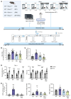
Figure 5. Germline Fbxw11 knockdown in male Nf1+/– mice attenuates cognitive deficits.
(A) The 4 genotypes of genetically engineered mice evaluated are shown. One group (n = 8 per genotype) performed the OF, CAR, and SP tasks. An independent cohort (n = 9 per genotype) performed the DDT due to the time-intensive training period. The indicated genotypes are abbreviated in parentheses for BE. Image created in BioRender. Angus, S. (2025) https://BioRender.com/g70k540 (B) Nf1+/– Fbxw11+/+ male mice exhibited hyperactive behavior in the OF test, with increased distance traveled over the 1-hour time period compared with WT Nf1+/+ Fbxw11+/+ mice (P = 0.0007, by 1-way ANOVA with Dunnett’s multiple-comparison test; mean ± SEM). For heterozygous Nf1 mutant mice (Nf1+/– Fbxw11+/+), Fbxw11 knockdown reduced hyperactive behavior to levels not significantly different from WT control mice. Fbxw11 had no adverse effects in WT Nf1+/+ Fbxw11+/–. Similar patterns of behavioral rescue were demonstrated in the DDT (C), SP (D), and CAR (E) tasks. (C) For the DDT, Nf1+/– Fbxw11+/+ mice showed increased small impulsive reward choices (including small and switch choices) rather than large delayed reward choices compared with WT animals (P = 0.0017, by 1-way ANOVA with Dunnett’s multiple-comparison test, mean ± SEM), and Nf1+/+ Fbxw11+/– mice showed no adverse behavioral effects. (D) In the SP task, we expected increased time spent with the novel mouse compared with the familiar mouse on day 1 and day 2 for the Nf1+/+ Fbxw11+/+ and Nf1+/+ Fbxw11+/– control groups. Nf1+/– Fbxw11+/+ mice showed decreased distinction between a novel partner mouse and a familiar partner mouse on day 2 (P = 0.1807), while Nf1+/– Fbxw11+/– mice showed a preference for the novel partner mouse on day 1 (P < 0.0001) and day 2 (P = 0.0010) by mixed-effects analysis and Tukey’s multiple-comparison test (mean ± SEM). (E) Although no differences by genotype were identified for edge entries (P = 0.0508), the number of falls (P < 0.0001), over-edge entries (P = 0.0006), and edge time (P = 0.0323) were increased for Nf1+/– Fbxw11+/+ mice compared with Nf1+/+ Fbxw11+/+, Nf1+/+ Fbxw11+/–, and Nf1+/– Fbxw11+/– animals (1-way ANOVA with Dunnett’s multiple-comparison test, mean ± SEM). *P < 0.05, **P < 0.01, ***P < 0.001, and ****P < 0.0001.
Figure 6
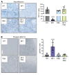
Figure 6. Germline Fbxw11 knockdown restores neurofibromin expression and suppresses ERK hyperactivation in Nf1+/– murine brain tissue.
IHC staining for (A) neurofibromin and (B) pERK1/2 in brain tissue from mice from Figure 5 was performed to determine the effect of heterozygous knockdown of Fbxw11, with representative images and quantification of immunoreactive cells shown. Original magnification, ×200. ****P < 0.0001, by 1-way ANOVA with Tukey’s multiple-comparison test. Data indicate the mean ± SEM.
Figure 7
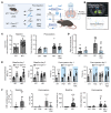
Figure 7. Selective deletion of Fbxw11 in the nucleus accumbens rescues cognitive deficits in male Nf1+/– Fbxw11fl/fl mice.
(A) Schematic of stereotaxic AAV virus injection. Cohorts of male Nf1+/– Fbxw11fl/fl and control Nf1+/+ Fbxw11fl/fl mice received bilateral injections of AAV-GFP (control, n = 8 mice per genotype) or AAV-Cre-GFP virus (n = 18 mice per genotype) for experiments (after injection). Mice underwent behavioral testing at baseline to confirm the expected deficits. The indicated genotypes and treatments are abbreviated as shown in parentheses for group labeling in CF. Image created in BioRender. Angus, S. (2025) https://BioRender.com/r74c640 (B) To ensure correct viral injection, IHC was performed to visualize GFP expression at the center of each injection site. A representative image of the injection site at the nucleus accumbens and center points of all injections is shown. (CF) Studies were performed as in Figure 4. (C) Nf1+/– Fbxw11fl/fl male mice exhibited hyperactivity in the OF task (P < 0.0001) at baseline and following GFP injection (P = 0.0053) compared with WT mice that was reduced in mice receiving CRE (P = 0.0056) as determined by 1-way ANOVA with Dunnett’s multiple-comparison test (mean ± SEM). (D) Nf1+/– Fbxw11fl/fl male mice infected with GFP made more impulsive choices in the DDT when compared with WT mice receiving GFP (P = 0.0001) that was suppressed in the CRE group. P = 0.0001, by 1-way ANOVA with Dunnett’s multiple-comparison test. Data indicate the mean ± SEM. (E) Nf1+/– Fbxw11fl/fl male mice spent more time investigating a novel partner on day 1 (P < 0.0001) but not day 2 (P = 0.1625) at baseline in the SP task. This pattern persisted in Nf1+/– Fbxw11fl/fl male mice receiving GFP, while CRE led to increased interaction time with a novel mouse versus a familiar mouse on day 2 (P < 0.0001). Significance was determined by mixed-effects analysis and Tukey’s multiple-comparison test. Data indicate the mean ± SEM. (F) Nf1+/– Fbxw11fl/fl male mice had a greater number of falls and over-the-edge time at baseline (P = 0.0009 and P < 0.0001, respectively) and after injection with GFP (P < 0.0001 and P = 0.0007, respectively) compared with WT mice in the CAR task. After CRE injection, the number of falls (P = 0.0003) and over-edge time (P = 0.002) for Nf1+/– Fbxw11fl/fl male mice was reduced compared with the GFP-injected cohort (NF1/FBX vs. NF1). Significance was determined by 1-way ANOVA with Dunnett’s multiple-comparison test. Data indicate the mean ± SEM. **P < 0.01, ***P < 0.001, and ****P < 0.0001. ##P < 0.01 and ###P < 0.001, for comparison of CRE-mediated Fbxw11 ablation with control GFP (NF1/FBX to NF1).
Figure 8
Figure 8. Targeted deletion of Fbxw11 in the nucleus accumbens of Nf1+/– mice corrects Nf1/Ras/MAPK pathway activity.
(A and B) IHC staining for neurofibromin (A) and pERK1/2 (B) was performed to determine the effect of targeted ablation of Fbxw11, with representative images and quantification of immunoreactive cells from mice as indicated in Figure 7. Original magnification, ×200. (C) Brain tissue from the nucleus accumbens region of mice of the indicated genotype and injected with the indicated AAV was used to generate lysate for RAF1 pull-down assays. Immunoblotting was used to detect vinculin (loading control) and Ras. *P < 0.05, **P < 0.01, and ****P < 0.0001. ####P < 0.0001, for comparison of CRE-mediated Fbxw11 ablation with control GFP (NF1/FBX to NF1). One-way ANOVA with Tukey’s multiple-comparison test. Data indicate the mean ± SEM.

References

    1. Gutmann DH, et al. Neurofibromatosis type 1. Nat Rev Dis Primers. 2017;3:17004. doi: 10.1038/nrdp.2017.4. - DOI - PubMed
    1. Hyman SL, et al. The nature and frequency of cognitive deficits in children with neurofibromatosis type 1. Neurology. 2005;65(7):1037–1044. doi: 10.1212/01.wnl.0000179303.72345.ce. - DOI - PubMed
    1. Ratner N, Miller SJ. A RASopathy gene commonly mutated in cancer: the neurofibromatosis type 1 tumour suppressor. Nat Rev Cancer. 2015;15(5):290–301. doi: 10.1038/nrc3911. - DOI - PMC - PubMed
    1. Diggs-Andrews KA, Gutmann DH. Modeling cognitive dysfunction in neurofibromatosis-1. Trends Neurosci. 2013;36(4):237–247. doi: 10.1016/j.tins.2012.12.002. - DOI - PMC - PubMed
    1. McQuade JD, Hoza B. Peer problems in attention deficit hyperactivity disorder: current status and future directions. Dev Disabil Res Rev. 2008;14(4):320–324. doi: 10.1002/ddrr.35. - DOI - PubMed