. 2025 Aug 4;22(8):4597-4611.
doi: 10.1021/acs.molpharmaceut.5c00125. Epub 2025 Jul 1.

Fabrication of RIG-I-Activating Nanoparticles for Intratumoral Immunotherapy via Flash Nanoprecipitation

Affiliations

Fabrication of RIG-I-Activating Nanoparticles for Intratumoral Immunotherapy via Flash Nanoprecipitation

Payton T Stone et al. Mol Pharm. .

Abstract

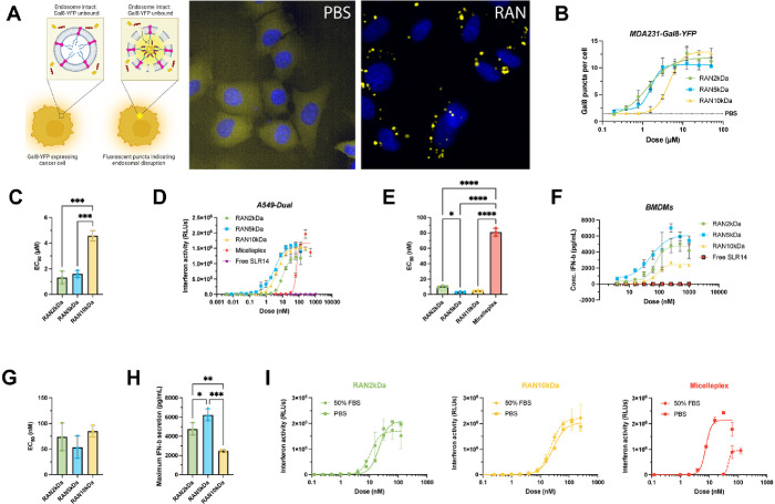
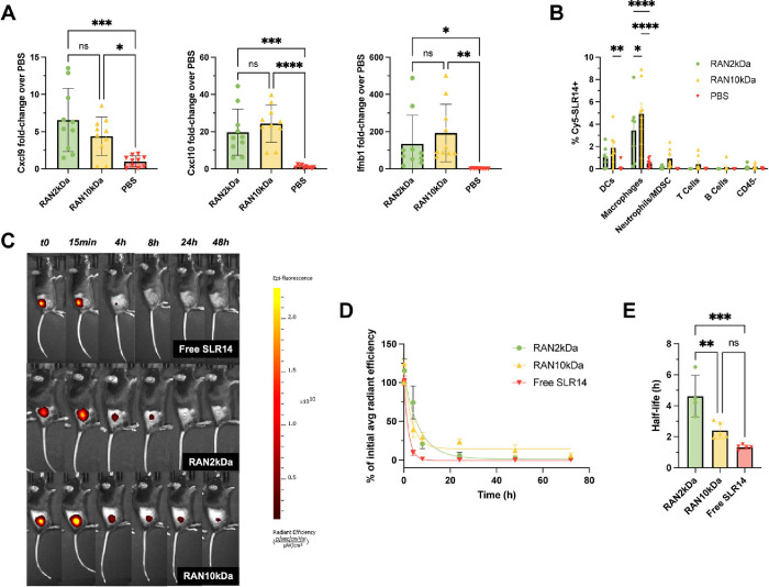
Intratumoral immunotherapy is a promising strategy for stimulating local and systemic antitumor immunity while eliminating or reducing immune-related adverse events often attendant to systemic administration. Activation of the cytosolic pattern recognition receptor retinoic acid-inducible gene I (RIG-I) at tumor sites stimulates innate immunity that can potentiate a T cell-dependent adaptive antitumor immune response. However, the activity and efficacy of 5'-triphosphate RNA (3pRNA) agonists of RIG-I are hindered by poor in vivo stability, rapid degradation, limited cellular uptake, and inefficient cytosolic delivery. To overcome these challenges, we developed RIG-I-activating nanoparticles (RANs) assembled using a flash nanoprecipitation (FNP) process to load a potent stem-loop 3pRNA (SLR) RIG-I agonist into endosome-destabilizing polymeric nanoparticles. We leveraged FNP to induce turbulent micromixing among a corona-forming poly(ethylene glycol)-block-(dimethylaminoethyl methacrylate-co-butyl methacrylate) (PEG-DB) diblock copolymer, a hydrophobic core-forming DB counterpart, and an SLR RIG-I agonist, resulting in the self-assembly of densely loaded nanoparticles that promoted endosomal escape and cytosolic delivery of 3pRNA cargo. Through optimization of polymer properties and inlet feed ratios, we developed RANs with high and improved loading efficiency and increased serum stability relative to a previously reported micelleplex formulation assembled via electrostatic complexation with PEG-DB polymers. We found that optimized RANs exhibited potent immunostimulatory activity in vitro and in vivo when delivered intratumorally. As a result, in preclinical models of MC38 colon cancer and B16.F10 melanoma, intratumoral administration of RANs suppressed tumor growth and increased survival time relative to vehicle controls. Collectively, this work demonstrates that FNP can be harnessed as a versatile and scalable process for the efficient loading of nucleic acids into polymeric nanoparticles and highlights the potential of RANs as a translationally promising platform for intralesional cancer immunotherapy.

Keywords: RIG-I; cancer immunotherapy; drug delivery; flash nanoprecipitation; nanoparticle; polymer.


Figures

1
1
Design, synthesis, and fabrication of RIG-I-activating nanoparticles (RANs). RAFT polymerization was used to synthesize pH-responsive, endosomolytic mPEG-block-[DMAEMA50%-co-BMA50%]6 kDa (PEG-DB) corona-forming diblock copolymers with mPEG molecular weights of 2, 5, and 10 kDa and a [DMAEMA-co-BMA]15 kDa (DB) core-forming copolymer (15 kDa) (created with ChemDraw 20.1.0.112). Flash nanoprecipitation (FNP) was employed to induce turbulent micromixing within a confined impingement jet (CIJ) mixer, facilitating particle self-assembly and encapsulation of 5′-triphosphorylated RNA (3pRNA) cargos within nanoparticles, resulting in the formation of RANs. After purification, RANs were administered intratumorally to stimulate an innate immune response that inhibited tumor growth and prolonged survival in murine cancer models. Upon administration, cells within the tumor microenvironment (TME) endocytose RANs, which disassemble and expose membrane lytic DB segments in response to a decrease in pH within the endosome. This culminates in endosomal disruption and subsequent release of triphosphorylated RNA into the cytosol where it can bind to and activate RIG-I to elicit antitumor innate immunity (created with Biorender.com).
2
2
Parameter optimization and characterization of RAN properties. (A) Initial screen for RAN size, polydispersity index (PDI), and encapsulation efficiency (EE) as a function of nitrogen to phosphate (N:P) ratio in the inlet CIJ streams (n = 2–5 experimental replicates per group) P values determined by an ordinary one-way ANOVA test with Tukey’s test for multiple comparisons. (B) Dynamic light scattering (DLS) intensity-weighted size distributions, (C) diameter, and (D) PDI for each RAN formulation (n = 4–5 experimental replicates per group). P values determined by an ordinary one-way ANOVA test with Dunnett’s test for multiple comparisons. (E) Representative transmission electron microscopy (TEM) images (scale bar = 50 nm) and (F) cryogenic electron microscopy (cryoEM) images (scale bar = 200 nm) of RAN2 kDa formulation. Assembly of RANs with FNP results in significantly higher (G) EEs and (H) loading capacities (LCs) than electrostatic complexation to form micelleplexes at the same N:P ratio of 8:1 (n = 4–5 experimental replicates per group). P values determined by an ordinary one-way ANOVA test with Dunnett’s test for multiple comparisons. (I) Zeta potential measurements (n = 2 experimental replicates per group). P values determined by an ordinary one-way ANOVA test with Tukey’s test for multiple comparisons. Replicates are experimental and technical, and data are shown as mean ± SD. **** signifies P < 0.0001.
3
3
RANs are endosomolytic, immunostimulatory in vitro, and stable in serum. (A) Experimental schematic of Gal8-YFP assay used to measure endosomolytic activity of RANs and representative microscopy images of MDA-MB-231 Gal8-YFP cells treated with a RAN formulation and a PBS control. (B) Dose response curves of MDA-MB-231 Gal8-YFP cells treated with indicated RAN formulations (n = 3 biological replicates per group). All RANs promoted endosomolytic activity in a dose-dependent manner as indicated by (C) calculated EC50 values of each RAN formulation. P values determined by an ordinary one-way ANOVA test with Tukey’s test for multiple comparisons. (D) Dose response curves for relative IFN-I production by A549-Dual reporter cells treated with RAN formulations, micelleplex, and free SLR14 controls (n = 3 biological replicates per group). (E) Calculated EC50 values for relative IFN-I production. P values determined by an ordinary one-way ANOVA test with Dunnett’s test for multiple comparisons. (F) Dose response curves for IFN-β secretion by isolated bone marrow-derived macrophages (BMDMs) treated with RAN formulations (n = 2 biological replicates per group). (G) Calculated EC50 values and (H) maximum secreted IFN-β levels. P values determined by an ordinary one-way ANOVA test with Tukey’s test for multiple comparisons. (I) Serum stability analysis of RAN2 kDa (green), RAN10 kDa (yellow), and micelleplex (red) immunostimulatory activity in A549-Dual IFN-I reporter cells after 24 h incubation in either 50% FBS or PBS at 37 °C. Replicates are biological, and data are shown as mean ± SD * signifies P < 0.05, ** signifies P < 0.01, *** signifies P < 0.001, **** signifies P < 0.0001.
4
4
RAN formulations are endocytosed by tumor-associated myeloid cells and stimulate innate immunity following intratumoral administration. (A) RAN formulations upregulate RIG-I-driven proinflammatory markers compared to PBS vehicle controls in MC38 tumors 4 h after one intratumoral treatment (n = 10 mice per group). P values determined by an ordinary one-way ANOVA test with Dunnett’s test for multiple comparisons. (B) Cellular uptake of Cy5-SLR14-OH cargo by indicated cell populations as determined by flow cytometry 4 h after intratumoral administration of RANs in MC38 tumor model (n = 7–9 mice per group). (C, D) Intratumorally administered RANs increase the retention time of SLR14 within the tumor vasculature compared to free drug. (E) RANs increase the half-life of SLR14 compared to free drug (n = 4–5 mice per group). P values determined by a two-way ANOVA test with Tukey’s test for multiple comparisons. Replicates are biological, and data are shown as mean ± SD * signifies P < 0.05, ** signifies P < 0.01, *** signifies P < 0.001, **** signifies P < 0.0001.
5
5
RANs mitigate disease progression and prolong survival in murine cancer models. (A) Experimental timeline and treatment schedule for intratumoral administration of mice with subcutaneous MC38 tumors. (B) Average tumor growth curves and (C) Kaplan–Meier survival plots for mice with MC38 tumors treated with indicated formulations. P values for tumor growth curves determined by a two-way ANOVA test with Tukey’s test for multiple comparisons on Day 14 shown. Survival curve comparisons were made using a Log-rank (Mantel-Cox) test. (D) Spider plots of individual tumor growth curves. (E) No notable weight loss was observed over the course of treatment (n = 7–9 mice per group). (F) Experimental timeline and treatment schedule for intratumoral administration of mice with subcutaneous B16.F10 tumors. (G) Average tumor growth curves and (H) Kaplan–Meier survival plots for mice with B16.F10 tumors treated with indicated formulations; CR = complete responder (n = 6–8 mice per group). P values determined by a two-way ANOVA test with Tukey’s test for multiple comparisons on Day 10 shown. Survival curve comparisons were made using a Log-rank (Mantel-Cox) test. Replicates are biological, and data are shown as mean ± SEM * signifies P < 0.05, ** signifies P < 0.01, **** signifies P < 0.0001.

Similar articles

References

    1. Jiang Y., Zhang H., Wang J., Chen J., Guo Z., Liu Y., Hua H.. Exploiting RIG-I-like Receptor Pathway for Cancer Immunotherapy. J. Hematol Oncol. 2023;16(1):8. doi: 10.1186/s13045-023-01405-9. - DOI - PMC - PubMed
    1. Kelly C. M., Antonescu C. R., Bowler T., Munhoz R., Chi P., Dickson M. A., Gounder M. M., Keohan M. L., Movva S., Dholakia R., Ahmad H., Biniakewitz M., Condy M., Phelan H., Callahan M., Wong P., Singer S., Ariyan C., Bartlett E. K., Crago A., Yoon S., Hwang S., Erinjeri J. P., Qin L.-X., Tap W. D., D’Angelo S. P.. Objective Response Rate Among Patients With Locally Advanced or Metastatic Sarcoma Treated With Talimogene Laherparepvec in Combination With Pembrolizumab. JAMA Oncol. 2020;6(3):402. doi: 10.1001/jamaoncol.2019.6152. - DOI - PMC - PubMed
    1. Toulmonde M., Cousin S., Kind M., Guegan J.-P., Bessede A., Le Loarer F., Perret R., Cantarel C., Bellera C., Italiano A.. Randomized Phase 2 Trial of Intravenous Oncolytic Virus JX-594 Combined with Low-Dose Cyclophosphamide in Patients with Advanced Soft-Tissue Sarcoma. J. Hematol Oncol. 2022;15(1):149. doi: 10.1186/s13045-022-01370-9. - DOI - PMC - PubMed
    1. Samson A., West E. J., Carmichael J., Scott K. J., Turnbull S., Kuszlewicz B., Dave R. V., Peckham-Cooper A., Tidswell E., Kingston J., Johnpulle M., da Silva B., Jennings V. A., Bendjama K., Stojkowitz N., Lusky M., Prasad K. R., Toogood G. J., Auer R., Bell J., Twelves C. J., Harrington K. J., Vile R. G., Pandha H., Errington-Mais F., Ralph C., Newton D. J., Anthoney A., Melcher A. A., Collinson F.. Neoadjuvant Intravenous Oncolytic Vaccinia Virus Therapy Promotes Anticancer Immunity in Patients. Cancer Immunol Res. 2022;10(6):745–756. doi: 10.1158/2326-6066.CIR-21-0171. - DOI - PMC - PubMed
    1. Mahalingam D., Wilkinson G. A., Eng K. H., Fields P., Raber P., Moseley J. L., Cheetham K., Coffey M., Nuovo G., Kalinski P., Zhang B., Arora S. P., Fountzilas C.. Pembrolizumab in Combination with the Oncolytic Virus Pelareorep and Chemotherapy in Patients with Advanced Pancreatic Adenocarcinoma: A Phase Ib Study. Clin. Cancer Res. 2020;26(1):71–81. doi: 10.1158/1078-0432.CCR-19-2078. - DOI - PMC - PubMed