. 2025 Dec;22(1):1-17.
doi: 10.1080/15476286.2025.2527494. Epub 2025 Jul 7.

SARS-CoV-2 RNA-binding protein suppresses extracellular miRNA release

Affiliations

SARS-CoV-2 RNA-binding protein suppresses extracellular miRNA release

Hyejin Mun et al. RNA Biol. 2025 Dec.

Abstract

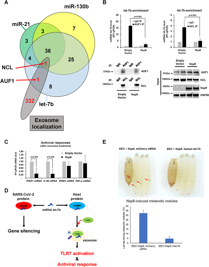
SARS-CoV-2 is the betacoronavirus causing the COVID-19 pandemic. Although the SARS-CoV-2 genome and transcriptome were reported previously, the function of individual viral proteins is largely unknown. Utilizing biochemical and molecular biology methods, we identified that four SARS-CoV-2 RNA-binding proteins (RBPs) regulate the host RNA metabolism by direct interaction with mature miRNA let-7b revealed by Nuclear Magnetic Resonance spectroscopy (NMR). SARS-CoV-2 RBP Nsp9 primarily binds mature miRNA let-7b, a direct ligand of the Toll-like Receptor 7 (TLR7), one of the potential SARS-CoV-2 therapeutics. Nsp9 suppresses host gene expression possibly by promoting let-7b-mediated silencing of a cellular RNA polymerase, POLR2D. In addition, Nsp9 inhibits extracellular release of let-7b and subsequent antiviral activity via TLR7. These results demonstrate that SARS-CoV-2 hijacks the host RNA metabolism to suppress antiviral responses and to shut down cellular transcription. Our findings of how a natural ligand of TLR7, miRNA let-7b, is suppressed by SARS-CoV-2 RBPs will advance our understanding of COVID-19 and SARS-CoV-2 therapeutics.

Keywords: Nsp9; POLR2D; SARS-CoV-2; let-7b; miRNA.


Conflict of interest statement

No potential conflict of interest was reported by the author(s).

Figures

Figure 1.
Figure 1.
SARS-CoV-2 Nsp9 promotes let-7b-mediated POLR2D mRNA decay.
Figure 2.
Figure 2.
SARS-CoV-2 Nsp9 interacts with let-7b.
Figure 3.
Figure 3.
Nsp9 and let-7b interaction analysis via 1H, 15N-HSQC NMR spectrum.
Figure 4.
Figure 4.
Observation of specific residues.
Figure 5.
Figure 5.
Structural analysis of Nsp9 upon let-7b interaction. (A) The RCI-S2 value of TALOS-N plot indicates the Nsp9 region’s flexibility (value near 0) and rigidity (value close to 1); (B) Chemical Shift Perturbation (CSP) illustrating the chemical shift differences between Nsp9 and Nsp9/let-7b complex, categorizing shifts into three regions, CSP > 0.06 ppm, CSP value between 0.03 ppm and 0.06 ppm, and CSP < 0.03 ppm. G4 and T22 are highlighted with stripes respectively, showcasing their unique spectral disappearances in the complex and isolated Nsp9; (C) Colour-coded dimer structure of Nsp9 based on CSP values: residues with CSPs > 0.06 ppm are depicted in red, while residues with CSPs between 0.03 ppm and 0.06 ppm are highlighted in yellow. G4 and T22 residues are uniquely coloured in violet and marine. N5, where the first N residue, is encircled by a dashed line. (D) Nsp9 phosphorylation structural models are presented, highlighting specific regions. Each Nsp9 dimer is depicted in cyan and yellow, with the N-terminal strand represented in magenta (-9 to 1, from the vector). The predicted phosphorylation site at residue T109 is marked in red within the alpha helices of each dimer. A distance measurement of 13.4 Å is indicated between the T109 phosphorylation sites of the dimer.
Figure 6.
Figure 6.
Nsp9 suppresses extracellular release of let-7b. (A-C) RT-qPCR levels of let-7b (A), miR-21 (B), and miR-130b (C) (normalized with U6 RNA) using exosomal RNAs purified from HeLa cells transfected with Strep-tag-Nsp9, Nsp13, Nsp14, Nsp15 plasmid or empty plasmid as a control for 48 h. Error bars represent the mean±SD of three independent experiments. *: p < 0.005. (D) Western blot analysis of the exosome marker protein CD63, the Golgi marker protein GM130, as well as AUF1 and NCL in the exosome pellet and supernatant (top). The size of each exosome was measured using nanoparticle tracking analysis, as indicated in the table (bottom).
Figure 7.
Figure 7.
Nsp9 suppresses exosomal let-7b release and TLR7-mediated antiviral responses. (A) Comparison of proteins binding with let-7b identified from protein microarrays, and enriched in exosomes from Mass Spectrometry of exosomal lysates. Two common RBPs, AUF1 and NCL, were highlighted. (B) RT-qPCR level of let-7b (normalized with U6 RNA) using RNAs pulled-down together with AUF1 (top, left) or NCL (top, right) from lysates of HeLa cells transfected with Strep-tag-Nsp9 plasmid or empty plasmid as a control for 48 h. Western blot analysis of the pull-down efficiency of AUF1 and NCL in Nsp9-overexpressing HeLa cells compared to control (bottom, left) and total cell lysates was probed with antibodies against AUF, NCL, Nsp9, and HSP90, as shown (bottom, right). Antibody against normal mouse IgG was used as background control. Error bars represent the mean±SD of three independent experiments. *: p < 0.005. (C) RT-qPCR level of TLR7-target mRNAs (normalized with GAPDH mRNA) using total RNAs purified from WI-38 cells exposed to exosomes for 2 h from HeLa cells transfected with Strep-tag-Nsp9 plasmid or empty plasmid as a control for 48 h. Error bars represent the mean±SD of three independent experiments. *: p < 0.005. (D) Proposed model of Nsp9 promoting let-7b-mediated gene silencing and suppressing extracellular let-7b release for antiviral responses. (E) Melanotic nodules in Nsp9-expressing larvae and pupae in the presence or absence of human let-7b (top). Quantification of larvae carrying melanotic nodules (bottom). More than 300 larvae were examined in three independent crosses. Error bars represent the mean±SD of three independent experiments. *p < 0.001.

References

    1. Zhu N, Zhang D, Wang W, et al. A novel coronavirus from patients with pneumonia in China, 2019. N Engl J Med. 2020;382(8):727–733. doi: 10.1056/NEJMoa2001017 - DOI - PMC - PubMed
    1. Zhou P, Yang X-L, Wang X-G, et al. A pneumonia outbreak associated with a new coronavirus of probable bat origin. Nature. 2020;579(7798):270–273. doi: 10.1038/s41586-020-2012-7 - DOI - PMC - PubMed
    1. Kim J-M, Chung Y-S, Jo HJ, et al. Identification of coronavirus isolated from a patient in Korea with COVID-19. Osong Public Health Res Perspect. 2020;11(1):3. doi: 10.24171/j.phrp.2020.11.1.02 - DOI - PMC - PubMed
    1. Menachery VD, Graham RL, Baric RS.. Jumping species—a mechanism for coronavirus persistence and survival. Curr Opin Virol. 2017;23:1–7. doi: 10.1016/j.coviro.2017.01.002 - DOI - PMC - PubMed
    1. Snijder E, Decroly E, Ziebuhr J.. The nonstructural proteins directing coronavirus RNA synthesis and processing. Adv Virus Res. 2016;96:59–126. - PMC - PubMed