

Motor network pre-habilitation by low-frequency repetitive transcranial magnetic stimulation. A proof-of-concept
- PMID: 40591056
- PMCID: PMC12214039
- DOI: 10.1007/s00701-025-06592-7
Motor network pre-habilitation by low-frequency repetitive transcranial magnetic stimulation. A proof-of-concept
Erratum in
-
Correction to: Motor network pre-habilitation by low-frequency repetitive transcranial magnetic stimulation. A proof-of-concept.Acta Neurochir (Wien). 2025 Jul 24;167(1):201. doi: 10.1007/s00701-025-06621-5. Acta Neurochir (Wien). 2025. PMID: 40702142 Free PMC article. No abstract available.
Abstract
Background: Tumors involving motor-eloquent brain regions pose a significant surgical challenge, as maximizing resection while preserving motor function requires a delicate balance. Neuromodulation-induced cortical prehabilitation (NICP) has emerged as a potential strategy to promote functional reorganization before surgery, potentially expanding the margins of safe resection.
Objective: This pilot study aimed to investigate whether accelerated, low-frequency repetitive transcranial magnetic stimulation (rTMS) targeting the right primary motor cortex (M1) could induce functional and microstructural changes in the motor network.
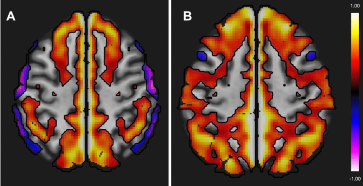
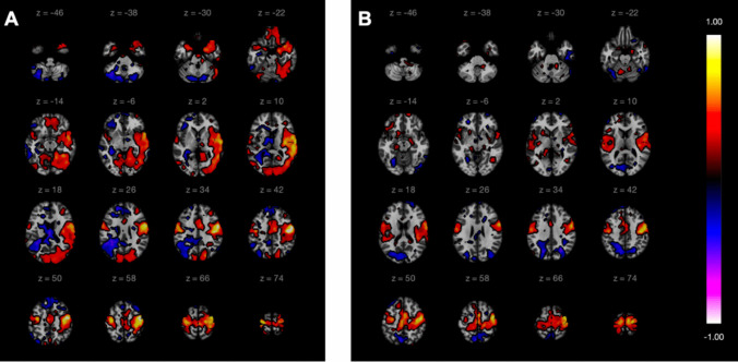
Methods: Two healthy subjects underwent a seven-day intervention consisting of twice-daily sessions of inhibitory rTMS over the right M1 (14 sessions in total). Pre- and post-intervention imaging included resting-state functional MRI (rs-fMRI) and diffusion tensor imaging (DTI). Functional changes were assessed descriptively using seed-based and ROI-to-ROI connectivity analyses. Microstructural changes were evaluated through tract-specific comparisons of fractional anisotropy (FA).
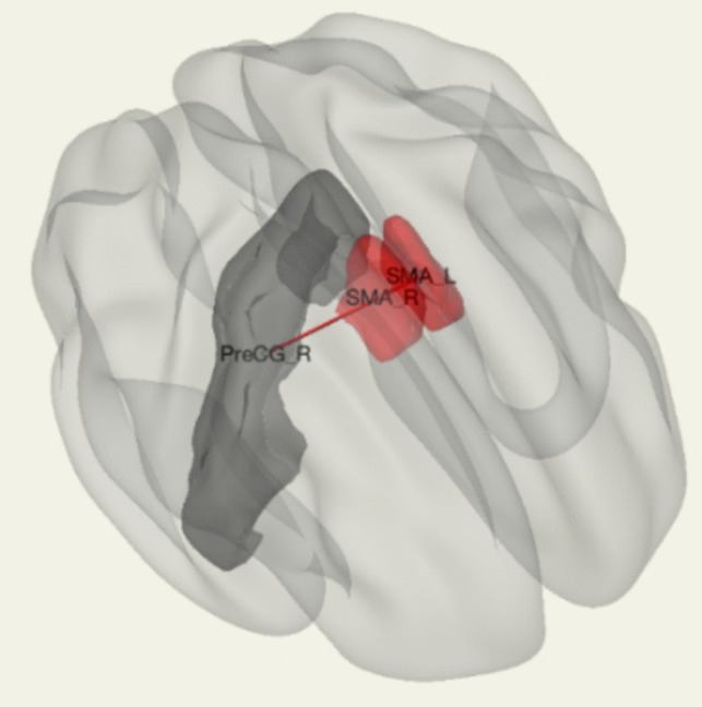
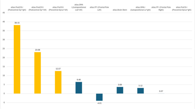
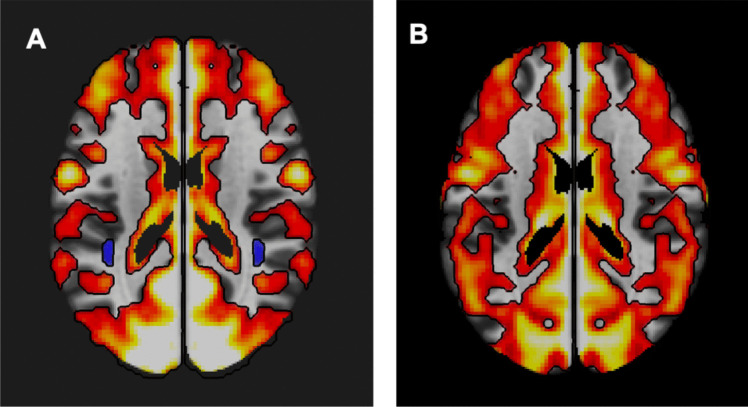
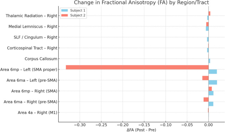
Results: Both subjects exhibited increased interhemispheric functional connectivity and strengthening of compensatory motor pathways, including the supplementary motor areas and bilateral precentral and postcentral gyri. DTI revealed tract-specific changes in FA, with evidence of microstructural modulation in regions such as the SMA, corpus callosum, and corticospinal tract. The magnitude and spatial distribution of changes varied between individuals.
Conclusion: These preliminary findings provide exploratory support for the hypothesis that inhibitory rTMS can induce functional and structural reorganization of the motor network. The combined use of rs-fMRI and DTI highlights the potential of NICP as a prehabilitation strategy in neurosurgical contexts. Further studies in clinical populations are warranted.
Keywords: Accelerated rTMS; Functional connectivity; Glioma; Neuromodulation; Neurorehabilitation; Neurosurgery; Plasticity; Prehabilitation; Sensorimotor Network; Transcranial magnetic stimulation.
© 2025. The Author(s).
Conflict of interest statement
Declarations. Informed consent: Written informed consent has been obtained from the patient to publish this paper. Institutional review board statement: The study was conducted in accordance with the Declaration of Helsinki, and approved by the Institutional Ethics Committee of CE-AVEC Azienda USL Bologna and Azienda USL Imola (protocol code N° 362–2024-OSS-AUSLBO approved on July 18th, 2024). Competing interests: The authors declare no competing interests.
Figures


Similar articles
-
The neuronal reserve in glioma surgery: functional reorganization of the motor network examined by navigated transcranial magnetic stimulation and diffusion tensor imaging tractography.J Neurosurg. 2025 May 2;143(3):793-804. doi: 10.3171/2025.1.JNS241103. Print 2025 Sep 1. J Neurosurg. 2025. PMID: 40315603
-
Application of non-invasive brain stimulation combined with functional magnetic resonance imaging in post-stroke motor function rehabilitation.J Neurosci Methods. 2025 Sep;421:110470. doi: 10.1016/j.jneumeth.2025.110470. Epub 2025 May 6. J Neurosci Methods. 2025. PMID: 40339709 Clinical Trial.
-
Advancing preoperative assessment of the motor system through navigated transcranial magnetic stimulation-based mapping of the supplementary motor area in patients with glioma.Neurosurg Focus. 2025 Aug 1;59(2):E7. doi: 10.3171/2025.5.FOCUS25298. Neurosurg Focus. 2025. PMID: 40749224
-
Impact of repetitive transcranial magnetic stimulation on cortical activity: a systematic review and meta-analysis utilizing functional near-infrared spectroscopy evaluation.J Neuroeng Rehabil. 2024 Jun 24;21(1):108. doi: 10.1186/s12984-024-01407-9. J Neuroeng Rehabil. 2024. PMID: 38915003 Free PMC article.
-
Non-invasive brain stimulation techniques for chronic pain.Cochrane Database Syst Rev. 2018 Mar 16;3(3):CD008208. doi: 10.1002/14651858.CD008208.pub4. Cochrane Database Syst Rev. 2018. Update in: Cochrane Database Syst Rev. 2018 Apr 13;4:CD008208. doi: 10.1002/14651858.CD008208.pub5. PMID: 29547226 Free PMC article. Updated.
References
-
- Amunts K, Mohlberg H, Bludau S, Zilles K (2020) Julich-Brain: a 3D probabilistic Atlas of the human brain’s cytoarchitecture. Science 369:988–992. 10.1126/science.abb4588 - PubMed
-
- Arumugham SS, Subhasini VS, Madhuri HN, Vinay B, Ravi M, Sharma E, Thirthalli J, Reddy YJ (2018) Augmentation Effect of Low-Frequency Repetitive Transcranial Magnetic Stimulation Over Presupplementary Motor Area in Obsessive-Compulsive Disorder: A Randomized Controlled Trial. J ECT 34:253–257. 10.1097/YCT.0000000000000509 - PubMed
-
- Barcia JA, Sanz A, Balugo P, Alonso-Lera P, Brin JR, Yus M, Gonzalez-Hidalgo M, Acedo VM, Oliviero A (2012) High-frequency cortical subdural stimulation enhanced plasticity in surgery of a tumor in Broca’s area. NeuroReport 23:304–309. 10.1097/WNR.0b013e3283513307 - PubMed
-
- Barcia JA, Sanz A, González-Hidalgo M, De Las Heras C, Alonso-Lera P, Díaz P, Pascual-Leone A, Oliviero A, Ortiz T (2012) rTMS Stimulation to induce plastic changes at the language motor area in a patient with a left recidivant brain tumor affecting Broca’s area. Neurocase 18:132–138. 10.1080/13554794.2011.568500 - PubMed
-
- Barwood CHS, Murdoch BE, Whelan B-M, Lloyd D, Riek S, O’Sullivan JD, Coulthard A, Wong A (2012) Improved receptive and expressive language abilities in nonfluent aphasic stroke patients after application of rTMS: an open protocol case series. Brain Stimul 5:274–286. 10.1016/j.brs.2011.03.005 - PubMed
MeSH terms
Grants and funding
LinkOut - more resources
Full Text Sources
