. 2025 Jul 1;16(1):1192.
doi: 10.1007/s12672-025-03021-0.

PDS5B positively regulates PTEN by binding to YTHDC1 to inhibit the malignant progression of endometrial carcinoma

Affiliations

PDS5B positively regulates PTEN by binding to YTHDC1 to inhibit the malignant progression of endometrial carcinoma

Hanyi Wang et al. Discov Oncol. .

Abstract

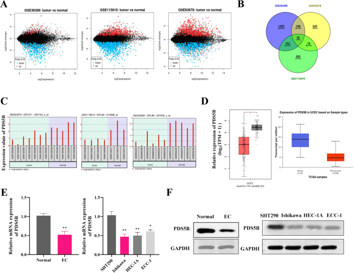
Background: Endometrial carcinoma (EC) is a common gynecological malignancy with a complex pathogenesis. PDS5B is revealed to be dysregulated and play critical roles in multiple cancers, while its role in EC remains largely unknown. The aim of this study was to explore the expression profile and biological function of the PDS5B in EC.

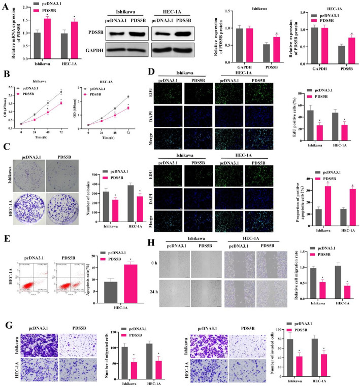
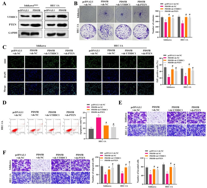
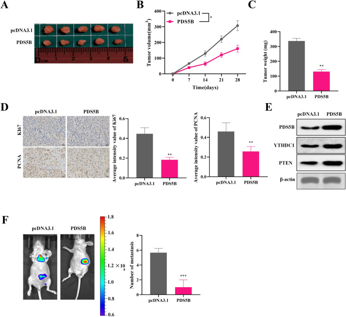
Methods: In this study, differential gene expression analysis in EC was performed using public databases and relevant analytical tools. The expression of PDS5B was verified by western blot and qRT-PCR in cell and tissue samples. The effects of PDS5B overexpression on EC cell proliferation, invasion and apoptosis, as well as the interaction of PDS5B with YTHDC1 and the regulation of PTEN stability were further explored by in vitro experiments. The effects of PDS5B on tumorigenesis and metastasis were explored using in vivo models.

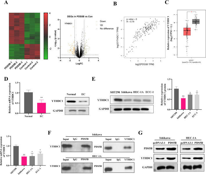
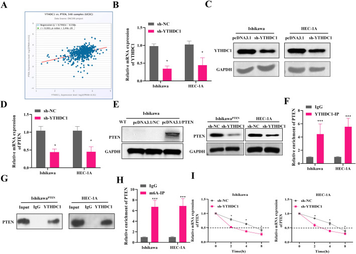
Results: PDS5B was down-regulated in EC, and overexpression of PDS5B significantly inhibited cell growth, and promoted apoptosis. In vitro results revealed that PDS5B interacted with YTHDC1 in EC cells and YTHDC1 regulated PTEN expression and stability via m6A modification, affecting the biological behavior of EC cells. In in vivo nude mouse models, overexpression of PDS5B significantly inhibited EC tumor development and metastasis in nude mice.

Conclusions: PDS5B interacts with YTHDC1 and further regulates PTEN in endometrial cancer cells, thereby inhibiting the malignant development of the tumor. These findings indicate the potential part of PDS5B in the development of EC and provide new molecular targets for the treatment of EC.

Keywords: Endometrial carcinoma; M6A; PDS5B; PTEN; YTHDC1.


Conflict of interest statement

Declarations. Ethics approval and consent to participate: This study was performed according to the principles of the Declaration of Helsinki, and was approved by the Joint Ethics Committee of the Ministry of Health. Informed consent was obtained from all individual participants included in the study. This study was approved by the Laboratory Animal Ethics Committee of Changzhou Geriatric Hospital Affiliated to Soochow University, Changzhou No. 7 People’s Hospital, and complied with the rules of Declaration of Helsinki principles. The maximal tumor size permitted by the ethics committee was 1000 mm3 and the maximal tumor burden was not exceeded in this study. Consent for publication: Not applicable. Competing interests: The authors declare no competing interests.

Figures

Fig. 1
Fig. 1
PDS5B expression is down-regulated in EC. A MA plots of differentially expressed genes in EC patients and normal patients in the GSE36389, GSE115810 and GSE63678 datasets in the GEO database. B Common differentially expressed genes in the three datasets. C Expression of PDS5B gene in the three datasets. D Expression of PDS5B gene in GEPIA and TCGA databases. E qRT-PCR and F western blot detection of PDS5B expression in clinical patient tissues (n = 3) and EC cell lines (n = 3). *p < 0.05, **p < 0.01
Fig. 2
Fig. 2
Low expression of PDS5B promotes proliferation and migration and inhibits apoptosis in EC. A qRT-PCR and western blot detection of PDS5B expression in Ishikawa and HEC-1 A cell line. B CCK-8 assay for cell viability. C Colony formation assay for cell proliferation. D EdU assay for cell proliferation. Bar scale = 100 μm. E TUNEL assay for cell apoptosis. Bar scale = 100 μm. F Flow cytometry for apoptosis. G Transwell assay for migration and invasion. H Wound healing assay for migration. *P < 0.05
Fig. 3
Fig. 3
PDS5B interacts with YTHDC1. A Schematic representation of RNA-seq of lshikawa cells overexpressing PDS5B or control. B GEPIA database showing PDS5B correlation with YTHDC1 in endometrial cancer. C GEPIA data showing the expression of YTHDC1 in endometrial cancers. qRT-PCR and western blot detection of YTHDC1 expression in EC D tissues and E cell lines. F Co-IP to detect the interaction of PDS5B with YTHDC1. G Western blot detection of YTHDC1 expression after overexpression of PDS5B. *p < 0.05, **p < 0.01
Fig. 4
Fig. 4
Targets of m6A modification of PTEN by YTHDC1 in EC. A The expression correlation between YTHDC1 and PTEN in UCEC in starBase database. B qRT-PCR and C western blot detection of YTHDC1 expression after YTHDC1 knockdown. D qRT-PCR detected PTEN mRNA expression after YTHDC1 knockdown. E Western blot detected PTEN protein expression in wild-type Ishikawa cells, Ishikawa cells transfected with pcDNA3.1/NC, or Ishikawa cells transfected with pcDNA3.1/PTEN for PTEN overexpression (IshikawaPTEN cells). Western blot also measured PTEN expression in IshikawaPTEN and HEC-1 A cells after YTHDC1 knockdown. F-G RIP to detect the interaction of YTHDC1 with PTEN. H MeRIP to detect m6A level of PTEN in cells. I Actinomycin D assay to detect the stability of PTEN after YTHDC1 knockdown. NC, negative control; *P < 0.05, ***P < 0.001
Fig. 5
Fig. 5
PDS5B inhibits malignant progression of EC through YTHDC1/PTEN. A Western blot detection of YTHDC1/PTEN expression after PDS5B overexpression. B Colony formation assay for cell proliferation. C Cell proliferation was detected by EdU. Bar scale = 100 μm. D Apoptosis was detected by flow cytometry. E-F Transwell assay for migration and invasion. *P < 0.05, *compared with pcDNA 3.1 + sh-NC, #P < 0.05, #compared with PDS5B + sh-NC
Fig. 6
Fig. 6
PDS5B inhibits EC cell growth and metastasis in vivo. A Schematic of mouse xenograft tumors (n = 5). B Tumor volume and C tumor mass. D IHC staining to analyze the protein expression levels of Ki67 and PCNA. Bar scale = 100 μm. E Western blot detection of PDS5B, YTHDC1 and PTEN expression in tumor tissue. F Monitoring in situ tumor metastasis by in vivo imaging system and H&E staining. **p < 0.01, ***p < 0.001

Similar articles

References

    1. Sakna NA, Elgendi M, Salama MH, et al. Diagnostic accuracy of endometrial sampling tests for detecting endometrial cancer: a systematic review and meta-analysis. BMJ Open. 2023;13(6):e072124. - PMC - PubMed
    1. Jónsdóttir B, Wikman A, Sundström Poromaa I, et al. Advanced gynecological cancer: quality of life one year after diagnosis. PLoS ONE. 2023;18(6):e0287562. - PMC - PubMed
    1. Siegel RL, Giaquinto AN, Jemal A. Cancer statistics, 2024. CA Cancer J Clin. 2024;74(1):12–49. - PubMed
    1. Xia C, Dong X, Li H, et al. Cancer statistics in China and united states, 2022: profiles, trends, and determinants. Chin Med J (Engl). 2022;135(5):584–90. - PMC - PubMed
    1. Colombo N, Creutzberg C, Amant F, et al. ESMO-ESGO-ESTRO consensus conference on endometrial cancer: diagnosis, treatment and Follow-up. Int J Gynecol Cancer. 2016;26(1):2–30. - PMC - PubMed

LinkOut - more resources