

Investigation of digital and conventional methods for verifying the fitness of CAD/CAM crowns on abutments with different shapes
- PMID: 40591199
- PMCID: PMC12214169
- DOI: 10.1186/s40729-025-00632-8
Investigation of digital and conventional methods for verifying the fitness of CAD/CAM crowns on abutments with different shapes
Abstract
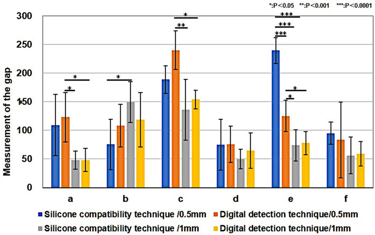
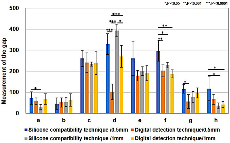
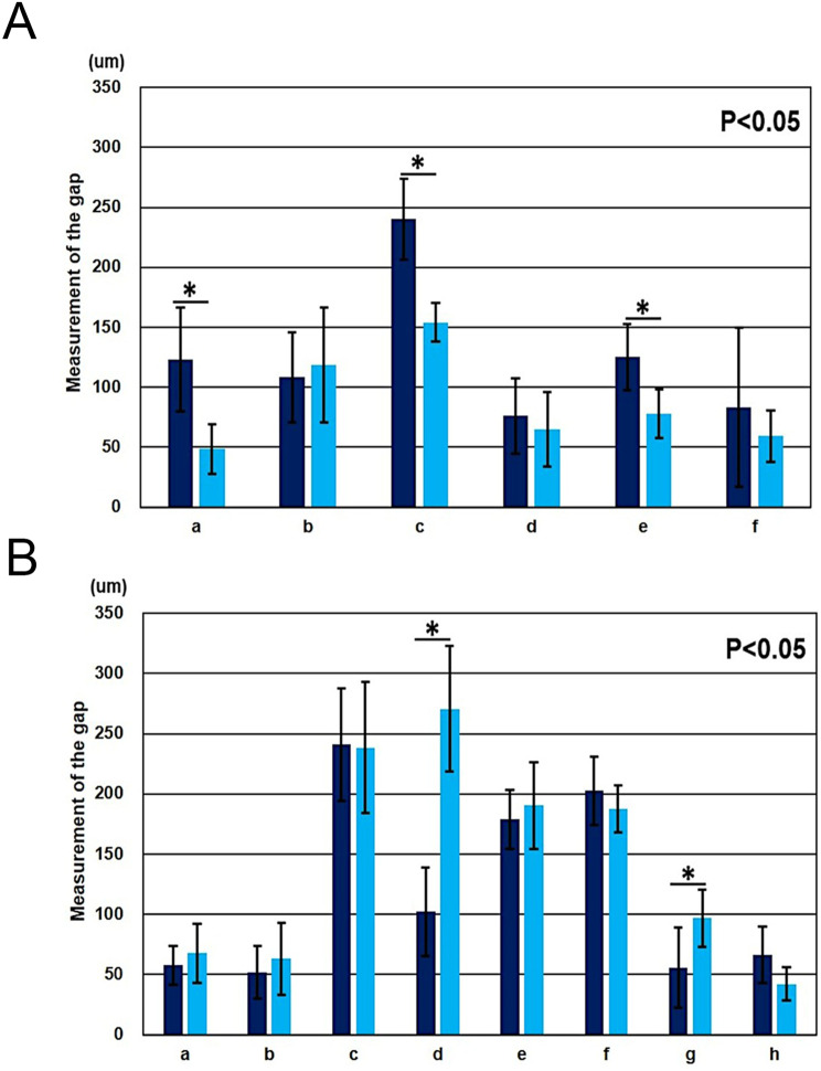
Purpose: To evaluate the marginal and internal compatibility of computer-aided design and computer-aided manufacturing crowns produced via a digital workflow using an intraoral scanner, and to compare this digital-detection technique with the conventional fit test using silicone rubber (silicone-compatibility technique) on various abutments.
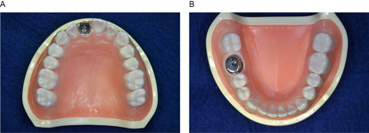
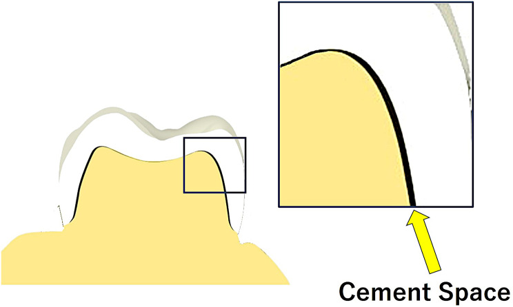
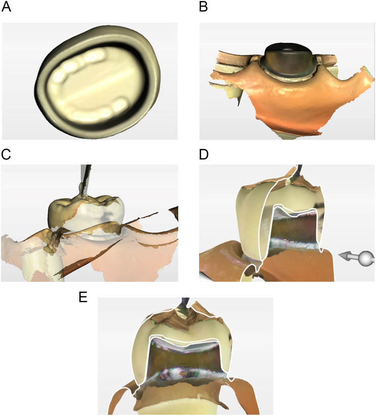
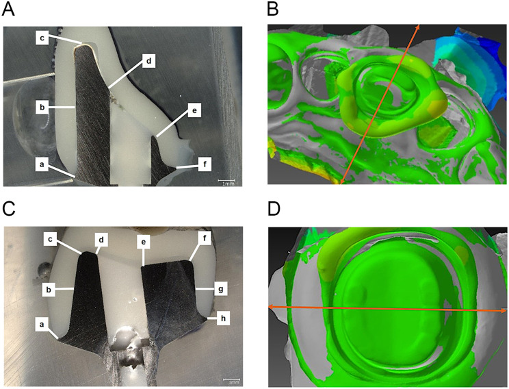
Methods: Implant bodies were placed in the maxillary right central incisor and mandibular right first molar of reference models. Digital scans were acquired using an intraoral scanner, and abutments were prepared. Twenty-four crowns with a cement space of 70 μm were fabricated from the digital file of the abutment. The crown's inner surface, abutment, and occlusal surface were scanned. The gaps between the crown and abutment were measured using stereoscopic image analysis software based on standard triangulated language data, and the accuracy of the fit was verified using silicone rubber.
Results: Significant differences (P < 0.05) were observed between the silicone-compatibility and digital-detection techniques for the maxillary central incisor at the incisal edge and the palatal lower region, and for the mandibular first molar at the occlusal surface and the center of lingual axis. The digital-detection technique yielded values closer to 70 μm for the cement space. The values measured using the silicone-compatibility technique exhibited greater variation than those measured using the digital-detection technique.
Conclusions: The novel digital-detection technique had superior or equivalent performance compared to the silicone-compatibility technique and could be beneficial for verifying crown fitness accuracy.
Keywords: CAD/CAM technology; Digital scan; Dimensional accuracy; Intraoral scan.
© 2025. The Author(s).
Conflict of interest statement
Declarations. Ethics approval and consent to participate: Not applicable. Consent for publication: Not applicable. Competing interests: The authors declare no competing interests.
Figures


Similar articles
-
Three-dimensional evaluation of the internal and marginal fit of multiunit implant-supported anterior fixed dental prostheses fabricated using different intraoral scanning protocols: An in vitro study.J Prosthet Dent. 2025 Sep;134(3):764.e1-764.e9. doi: 10.1016/j.prosdent.2025.06.001. Epub 2025 Jul 1. J Prosthet Dent. 2025. PMID: 40592691
-
Internal and marginal fit of digitally fabricated all-ceramic crowns with auxiliary retentive features on short clinical abutments: a micro-CT study.PeerJ. 2025 Aug 25;13:e19813. doi: 10.7717/peerj.19813. eCollection 2025. PeerJ. 2025. PMID: 40895059 Free PMC article.
-
Verification of a digital approach for three-dimensional evaluation of marginal and internal fit.J Prosthet Dent. 2025 Jul;134(1):160-166. doi: 10.1016/j.prosdent.2023.09.004. Epub 2023 Oct 17. J Prosthet Dent. 2025. PMID: 37852857
-
The complete digital workflow in fixed prosthodontics: a systematic review.BMC Oral Health. 2017 Sep 19;17(1):124. doi: 10.1186/s12903-017-0415-0. BMC Oral Health. 2017. PMID: 28927393 Free PMC article.
-
Marginal adaptation and CAD-CAM technology: A systematic review of restorative material and fabrication techniques.J Prosthet Dent. 2018 Apr;119(4):545-551. doi: 10.1016/j.prosdent.2017.07.001. Epub 2017 Sep 28. J Prosthet Dent. 2018. PMID: 28967399
References
-
- Arezoobakhsh A, Shayegh SS, Jamali Ghomi A, Hakimaneh SM. Comparison of marginal and internal fit of 3-unit zirconia frameworks fabricated with CAD-CAM technology using direct and indirect digital scans. J Prosthet Dent. 2020;123:105–12. - PubMed
-
- Miyazaki T, Hotta Y. CAD/CAM systems available for the fabrication of crown and Bridge restorations. Aust Dent J. 2011;56(Suppl 1):97–106. - PubMed
-
- Güth JF, Keul C, Stimmelmayr M, Beuer F, Edelhoff D. Accuracy of digital models obtained by direct and indirect data capturing. Clin Oral Investig. 2013;17:1201–8. - PubMed
-
- Brawek PK, Wolfart S, Endres L, Kirsten A, Reich S. The clinical accuracy of single crowns exclusively fabricated by digital workflow-the comparison of two systems. Clin Oral Investig. 2013;17:2119–25. - PubMed
-
- Nayyar N, Yilmaz B, McGlumphy E. Using digitally coded healing abutments and an intraoral scanner to fabricate implant-supported, cement-retained restorations. J Prosthet Dent. 2013;109:210–5. - PubMed
MeSH terms
LinkOut - more resources
Full Text Sources
Miscellaneous
