

doi: 10.1002/ctm2.70390.
Clinical translation potential of self-inspired live-cell super-resolution microscopy
Affiliations
- PMID: 40591244
- PMCID: PMC12211969
- DOI: 10.1002/ctm2.70390
Item in Clipboard
Clinical translation potential of self-inspired live-cell super-resolution microscopy
Clin Transl Med.
2025 Jul.
No abstract available
Keywords: clinical translation; live‐cell imaging; self‐supervised learning; super‐resolution microscopy.
Figures
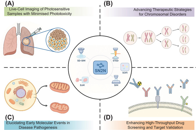
Clinical translational potential of the Self‐inspired Noise2Noise (SN2N)‐enhanced live‐cell super‐resolution microscopy. The SN2N framework (centre) is compatible with various super‐resolution microscopy modalities (e.g., spinning‐disc confocal‐based structured illumination microscopy [SD‐SIM], stimulated emission depletion [STED], SR optical fluctuation imaging reconstruction [SOFI], structured illumination microscopy [SIM], expansion microscopy [ExM]). By improving photon efficiency by one to two orders of magnitude, SN2N enables high‐fidelity imaging under reduced illumination, minimising phototoxicity and photobleaching. This facilitates diverse clinical research applications, including: (A) Live‐cell imaging of photosensitive samples (e.g., patient‐derived biopsies) with minimised phototoxicity for long‐term observation of disease progression and therapeutic responses. (B) Enabling extended, high‐resolution observation of dynamic processes like mitosis, offering insights into chromosomal instability mechanisms relevant to therapeutic strategies for chromosomal disorders. (C) Visualising subtle morphological and dynamic alterations for elucidating early diagnostic markers in diseases. (D) Improving high‐throughput drug screening and target validation by allowing accurate assessment of compound effects on cellular architecture and biomolecular interactions with minimal phototoxic perturbation. Schematics were created with BioRender.com.
Similar articles
-
Self-inspired learning for denoising live-cell super-resolution microscopy.Nat Methods. 2024 Oct;21(10):1895-1908. doi: 10.1038/s41592-024-02400-9. Epub 2024 Sep 11. Nat Methods. 2024. PMID: 39261639
-
Super-beacons: Open-source probes with spontaneous tuneable blinking compatible with live-cell super-resolution microscopy.Traffic. 2020 May;21(5):375-385. doi: 10.1111/tra.12728. Epub 2020 Mar 30. Traffic. 2020. PMID: 32170988 Free PMC article.
-
Rationalized deep learning super-resolution microscopy for sustained live imaging of rapid subcellular processes.Nat Biotechnol. 2023 Mar;41(3):367-377. doi: 10.1038/s41587-022-01471-3. Epub 2022 Oct 6. Nat Biotechnol. 2023. PMID: 36203012
-
Organic fluorescent probes for live-cell super-resolution imaging.Front Optoelectron. 2023 Nov 10;16(1):34. doi: 10.1007/s12200-023-00090-3. Front Optoelectron. 2023. PMID: 37946039 Free PMC article. Review.
-
Super-resolution radial fluctuations (SRRF): a versatile and accessible tool for live-cell nanoscopy.Histochem Cell Biol. 2025 Jun 18;163(1):67. doi: 10.1007/s00418-025-02396-z. Histochem Cell Biol. 2025. PMID: 40531238 Review.
References
Grants and funding
- 2022YFC3400600/National Key Research and Development Program of China
- 2024YFC3406600/National Key Research and Development Program of China
- 32422052/National Natural Science Foundation of China
- 62305083/National Natural Science Foundation of China
- T2222009/National Natural Science Foundation of China
- 32227802/National Natural Science Foundation of China
- 624B2054/National Natural Science Foundation of China
- 2023QNRC001/Young Elite Scientists Sponsorship Program by China Association for Science and Technology
- ZYGXQNJSKYCXNLZCXM-H7/Scientific Research Innovation Capability Support Project for Young Faculty
LinkOut - more resources
Full Text Sources