. 2025 Jul 1;20(7):e0327085.
doi: 10.1371/journal.pone.0327085. eCollection 2025.

Cl-amidine confers organ protection and improves survival in hemorrhagic shock rats via the PAD4-CitH3-NETs axis

Affiliations

Cl-amidine confers organ protection and improves survival in hemorrhagic shock rats via the PAD4-CitH3-NETs axis

Huiting Yun et al. PLoS One. .

Abstract

Background: The occurrence of multi-organ dysfunction following hemorrhagic shock (HS) remains a critical clinical challenge. The excessive formation of neutrophil extracellular trap (NET) Has been identified as a pivotal pathogenic mechanism. This study preliminarily elucidated the protective mechanism of the PAD4 inhibitor Cl-amidine in a rat model of HS.

Methods: Male Sprague-Dawley rats were subjected to sublethal (40% blood loss, n = 8) or lethal (50% blood loss, n = 10) HS. Rats were divided into Sham group (catheter placement only), HS group (catheter placement followed by blood withdrawal), Vehicle group (0.9% saline), and Cl-amidine (10 mg/kg in 0.9% saline) groups.

Results: Cl-amidine significantly improved the 72-h survival rate and delayed mortality in lethal HS. In Sublethal HS, the drug corrected metabolic disturbances, such as reduced lactate accumulation, while maintaining mean arterial pressure. Mechanistically, the effects of Cl-amidine included reducing circulating cell-free DNA (cf-DNA) and tissue citrullinated histone H3 (CitH3) levels, suppressing PAD4 expression, and improving histopathological outcomes (reduced edema and restored intestinal barrier integrity by upregulation of tight junction proteins Claudin-1/ZO-1). Moreover, Cl-amidine inhibited neutrophil infiltration through ICAM-1 downregulation and reduced the production of TNF-α and IL-6.

Conclusions: In conclusion, Cl-amidine protects against HS by targeting the PAD4-CitH3-NETs axis, breaking the vicious cycle of "NETs-inflammation", restoring barrier integrity, and alleviating multi-organ damage. The synergistic downregulation of ICAM-1 further enhances the therapeutic efficacy, highlighting Cl-amidine as a novel NETs-modulating strategy for HS. This study provides a theoretical and therapeutic foundation for the prevention and treatment of multi-organ injury following HS.


Conflict of interest statement

The authors have declared that no competing interests exis.

Figures

Fig 1
Fig 1. Experimental protocol and therapeutic outcomes of Cl-amidine in hemorrhagic shock.
(A) Experimental flowchart, (B) Kaplan-Meier survival curves were generated for all rat groups (n = 10 per group), and intergroup differences were analyzed using the log-rank test, (C) Changes in MAP curves of rats in each group (n = 8), and data are expressed as mean ± SD. Statistical significance was denoted as follows **p < 0.01, ***p < 0.001 vs. Sham group; ##p < 0.01, ###p < 0.001 vs. HS group; &p < 0.05, &&p < 0.01 vs. Vehicle group.
Fig 2
Fig 2. Effect of Cl-amidine on arterial blood gas in rats with hemorrhagic shock.
(A) Lactate (LAC, n = 8), (B) pH value (pH, n = 8), (C) Bicarbonate ion (HCO₃ ⁻ , n = 7), (D) Base excess (BE, n = 8), (E) Partial pressure of arterial oxygen (PaO₂, n = 8), (F) Partial pressure of arterial carbon dioxide (PaCO₂, n = 8), (G) P50 oxygen tension (n = 6), (H) Arterial oxygen saturation (SaO₂, n = 8). Data are expressed as mean ± SD, Statistical significance was denoted as follows *p < 0.05, **p < 0.01, ***p < 0.001 vs Sham; #p < 0.05, ###p < 0.001 vs HS; &p < 0.05, &&p < 0.01, &&&p < 0.001 vs Vehicle.
Fig 3
Fig 3. Effect of Cl-amidine on intestinal and lung tissue injury in rats with hemorrhagic shock.
(A) H&E-stained ileal sections (n = 4, scale bar: 50 µm): Black arrows: Separation of the epithelial apex from the lamina propria; Blue arrows: Swelling of villous epithelial cells; Red arrows: Focal neutrophil infiltration; Brown arrows: Nuclear fragmentation of epithelial cells; Cyan arrows: Lamina propria detachment. (B) Pathological injury scores of intestine (n = 4). (C) Wet/dry weight ratio of ileal tissues (n = 8). (D) H&E-stained pulmonary sections (n = 4, scale bar: 50 µm): Blue arrows: Widened alveolar septa with interstitial edema; Red arrows: Focal neutrophil infiltration; Black arrows: Thickened and disrupted alveolar walls; Brown arrows: Sloughing of pulmonary epithelial cells; Green arrows: Alveolar hemorrhage; Cyan arrows: Leukocyte margination. (E) Pathological injury scores of pulmonary tissues (n = 4). (F) Wet/dry weight ratio of Lung (n = 8). (G) Expression levels of Claudin-1 in intestinal tissue. (H) Grayscale value analysis of Claudin-1 expression levels in intestinal tissue. (I) Expression levels of ZO-1 in intestinal tissue and corresponding grayscale value analysis. (J) Grayscale value analysis of ZO-1 expression levels in intestinal tissue. Data are presented as mean ± SD. Statistical significance was denoted as follows *p < 0.05, **p < 0.01, ***p < 0.001 vs. Sham group; #p < 0.05, ##p < 0.01 vs. HS group; &p < 0.05, &&p < 0.01 vs. Vehicle group.
Fig 4
Fig 4. The effects of Cl-amidine on epithelial activation-induced neutrophils and inflammatory responses.
(A) Expression levels of ICAM-1 in intestinal tissue at 6 hours in different groups (n = 5). (B) Expression levels of CitH3 and grayscale value analysis in intestinal tissue at 6 hours in different groups. (C) Representative fluorescent staining images of NE and MPO in ileum tissue (n = 4, 200 × , scale bar: 50 µm). Fluorescence channels: NE (red), MPO (green), DAPI (nucleus, blue). (D) Percentage of MPO/ NE co-positive area in intestinal tissue. (E) Percentage of MPO/ NE co-positive cells in intestinal tissue. (F) TNF-α levels in intestinal tissue (n = 6). (G) IL-6 levels in intestinal tissue (n = 6). (H) IL-10 levels in intestinal tissue (n = 6). Data are presented as mean ± SD. Significant differences: *p < 0.05, **p < 0.01, ***p < 0.001 vs. Sham group; #p < 0.05, ##p < 0.01, ###p < 0.001 vs. HS group; &p < 0.05, &&p < 0.01, &&&p < 0.001 vs. Vehicle group.
Fig 5
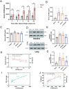
Fig 5. Effect of Cl-amidine on circulating and tissue NETs markers in rats with hemorrhagic shock and correlation analysis between NETs leve is and intestinal tissue injury.
(A) Changes in serum cf-DNA levels within 24 hours in rats with hemorrhagic shock (n = 4). (B) Serum cf-DNA levels at 6 hours in different groups (n = 8). (C) Serum CitH3 levels at 6 hours in different groups (n = 8). (D) Expression levels of CitH3 in intestinal tissue at 6 hours in different groups (n = 5). (E) Grayscale value analysis of CitH3 expression levels in intestinal tissue at 6 hours in different groups. (F) Expression levels of CitH3 in lung tissue at 6 hours in different groups (n = 5). (G) Grayscale value analysis of CitH3 expression levels in lung tissue at 6 hours in different groups. Data are presented as mean ± SD. Significant differences: * p < 0.05, ** p < 0.01 vs. sham group; # p < 0.05, ## p < 0.01 vs. HS group; & p < 0.05, &&p < 0.01 vs. vehicle group. (H) Correlation analysis between CitH3 expression levels and Claudin-1/ZO-1 expression levels in intestinal tissue using Spearman’s method. (I) Correlation analysis between serum cf-DNA and intestinal tissue wet/dry (W/D) weight ratio using Spearman’s method. (J) Correlation analysis between serum cf-DNA and intestinal tissue IL-6/TNF-α levels using Spearman’s method.
Fig 6
Fig 6. Cl-amidine inhibits PAD4-induced NETs levels.
(A) Quantitative real-time PCR analysis of PAD4 mRNA expression in intestinal tissue (n = 5). (B) Representative Western blot images of PAD4 protein expression in intestinal tissue (n = 5).(C) Quantitative analysis of PAD4 protein expression in intestinal tissue. (D) Representative immunofluorescence images of CitH3 and NE in intestinal tissue (n = 4, magnification 200 × , scale bar: 50 µm). Fluorescence channels: NE (red), MPO (green), DAPI (nucleus, blue). (E) Percentage of CitH3/NE co-positive cells in intestinal tissue. (F) Percentage of CitH3/NE co-positive area in intestinal tissue. Data are presented as mean ± SD. Significant differences: *p < 0.05, **p < 0.01, ***p < 0.001 vs. Sham group; #p < 0.05, ##p < 0.01 vs. HS group; &p < 0.05, &&p < 0.01 vs. Vehicle group.

Similar articles

References

    1. Cannon JW. Hemorrhagic shock. N Engl J Med. 2018;378(19):1850–3. doi: 10.1056/nejmc1802361 - DOI - PubMed
    1. Ali M, Pham A, Wang X, Wolfram J, Pham S. Extracellular vesicles for treatment of solid organ ischemia-reperfusion injury. Am J Transplant. 2020;20(12):3294–307. doi: 10.1111/ajt.16164 - DOI - PubMed
    1. Pantalone D, Bergamini C, Martellucci J, Alemanno G, Bruscino A, Maltinti G, et al. The role of DAMPS in burns and hemorrhagic shock immune response: pathophysiology and clinical issues. review. Int J Mol Sci. 2021;22(13):7020. doi: 10.3390/ijms22137020 - DOI - PMC - PubMed
    1. Brockmann L, Tran A, Huang Y, Edwards M, Ronda C, Wang HH, et al. Intestinal microbiota-specific Th17 cells possess regulatory properties and suppress effector T cells via c-MAF and IL-10. Immunity. 2023;56(12):2719–35. doi: 10.1016/j.immuni.2023.11.003 - DOI - PMC - PubMed
    1. van der Poll T, Shankar-Hari M, Wiersinga WJ. The immunology of sepsis. Immunity. 2021;54(11):2450–64. doi: 10.1016/j.immuni.2021.10.012 - DOI - PubMed

MeSH terms

Substances

LinkOut - more resources