. 2025 Aug:118:105826.
doi: 10.1016/j.ebiom.2025.105826. Epub 2025 Jun 30.

Trajectory of plasma lipidome associated with the risk of late-onset Alzheimer's disease: a longitudinal cohort study

Affiliations

Trajectory of plasma lipidome associated with the risk of late-onset Alzheimer's disease: a longitudinal cohort study

Tingting Wang et al. EBioMedicine. 2025 Aug.

Abstract

Background: Comprehensive lipidomic studies have demonstrated strong cross-sectional associations between the blood lipidome and late-onset Alzheimer's disease (AD) dementia and its risk factors, yet the longitudinal relationship between lipidome changes and AD progression remains unclear.

Methods: We employed longitudinal lipidomic profiling on 4730 plasma samples from 1517 participants of the Alzheimer's Disease Neuroimaging Initiative (ADNI) cohort to investigate the temporal evolution of lipidomes among diagnostic groups. At baseline (n = 1393), participants were classified as stable diagnosis status including stable AD (n = 243), stable cognitive normal (CN; n = 337), and stable mild cognitive impairment (MCI; n = 413), or converters (AD converters: n = 329; MCI converters: n = 71). We developed a dementia risk classification model to stratify the non-converting MCI group into dementia-like and non-dementia-like MCI based on their baseline lipidomic profiles, aiming to identify early metabolic signatures predictive of dementia progression.

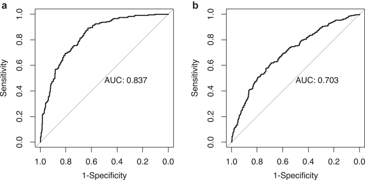
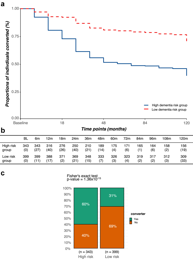
Findings: Longitudinal analysis identified significant associations between the change in ether lipid species (including alkylphosphatidylcholine, alkenylphosphatidylcholine, lysoalkylphosphatidylcholine, and lysoalkenylphosphatidylcholine) and AD dementia conversion. Specifically, AD dementia converters show a 3-4.8% reduction in these ether lipid species compared to the non-converting CN and MCI groups, suggesting metabolic dysregulation as a key feature of AD progression. Further, The Dementia Risk Model effectively distinguished MCI from AD dementia converters (AUC = 0.70; 95% CI: 0.66-0.74). Within the MCI group, the model identified a high-risk subgroup with a twofold higher likelihood of conversion to AD dementia compared to the low-risk group. External validation in the ASPREE cohort confirmed its predictive utility, with the Dementia Risk Score discriminating incident dementia from cognitively normal individuals (C-index = 0.75, 95% CI: 0.73-0.78), improving prediction by 2% over the combination of traditional risk factors and APOE genetic risk factor. Additionally, the Dementia Risk Score was significantly associated with reduced temporal lobar fludeoxyglucose uptake (β = -0.286, p = 1.34 × 10⁻⁴), higher amyloid PET levels (β = 0.308, p = 4.03 × 10⁻⁴), and elevated p-tau levels (β = 0.167, p = 2.37 × 10⁻²), reinforcing its pathophysiological relevance in tracking neurodegeneration, amyloid burden, and tau pathology.

Interpretation: These findings highlight lipidomic profiling as a potential blood-based biomarker for identifying individuals at high risk of AD progression, offering a scalable, non-invasive approach for early detection, risk stratification, and targeted interventions in AD.

Funding: The National Health and Medical Research Council of Australia (#1101320 and #1157607); NHMRC Investigator grant (#GNT1197190); Victorian Government's Operational Infrastructure Support Program; National Heart Foundation of Australia, Future Leader Fellowship (#102604), and National Health and Medical Research Council Investigator Grant (#2026325); Investigator grant (#2009965) from the National Health and Medical Research Council of Australia; a National Health and Medical Research Council of Australia Senior Research Fellowship (#1042095); National Institutes of Health grants: P30AG010133, P30AG072976, R01AG019771, R01AG057739, U19AG024904, R01LM013463, R01AG068193, T32AG071444, U01AG068057, U01AG072177, U19AG074879, R01AG069901, R01AG046171, RF1AG051550, RF1AG057452; National Institutes of Health/National Institute on Aging grants RF1AG058942, RF1AG059093, U01AG061359, U19AG063744, and R01AG081322, NIH/NLM R01LM012535; FNIH: DAOU16AMPA.

Keywords: AD biomarkers; Alzheimer's disease; Cognitively normal; Lipidomics; Mild cognitive impairment.


Conflict of interest statement

Declaration of interests Dr. Kaddurah-Daouk is an inventor on a series of patents on use of metabolomics for the diagnosis and treatment of CNS diseases and holds equity in Metabolon Inc., Chymia LLC and PsyProtix. Prof. Meikle leads the provisional patent “METHODS OF ASSESSING ALZHEIMER’S DISEASE” on the development of dementia risk scores that has been filed with the Serial No. 63/463,808. Gabi Kastenmüller declares equity in Chymia LLC and EMBL-EBI, is the inventor on patents, Philippine Genome Center/Davao Medical School, DAAD/DWIH, and BMBF. Andrew Saykin has received gifts/services from Avid Radiopharmaceuticals, has editorial involvement with Springer-Nature Publishing, and has served on scientific advisory boards for Eisai, Novo Nordisk, and Siemens. Paul Lacaze is supported by a National Heart Foundation of Australia Future Leader Fellowship. Matthias Arnold reports support from NIH/NIA, is a co-inventor on patents, and has equity in Chymia LLC, PsyProtiz, and Atai Life Sciences. Other authors have declared that no conflict of interest exists.

Figures

Fig. 1
Fig. 1
Study design. This study had two parts. Part 1 involved the development of a Dementia risk model, using baseline data, to characterise the heterogeneity of the non-converting MCI group and to calculate lipidomic risk scores for individuals across different time points. A ridge regression model, built within a five-fold cross-validation framework was used to stratify the non-converting MCI group into dementia-like and non-dementia-like sub-groups. In the development of the model, we treated Dementia risk status as outcome with the predictors including all the lipid species, age, sex, BMI, clinical lipids, fasting status, cohort, APOE ε4, omega-3, and statin status. Part 2 was the longitudinal analysis on the repeated measurements across 13 time points to examine the associations of changes in lipid species and lipidomic risk scores with AD dementia status. Associations of the trajectories of individual lipid species and disease outcomes were examined using linear mixed models to undercover the difference of trajectories of lipid species between different groups. The covariates included age, sex, BMI, fasting status, cohort, APOE ε4, omega-3, and statin status. Thereafter, global lipidomic scores combing all lipid species derived from the Dementia risk model were fitted into a linear mixed model to define the associations with AD related biomarkers. Similarly, the covariate set included age, sex, BMI, clinical lipids, fasting status, cohort, APOE ε4, omega-3, and statin status.
Fig. 2
Fig. 2
Trajectory of lipid species between different AD dementia diagnosis groups across all the time points. The linear mixed model was performed to examine the association of the changes of individual lipid species with AD dementia diagnosis state. a) After excluding AD dementia cases, we compared the converters (n = 363 with 1353 repeated measurements) vs non-converter groups (the combination of non-converting CN and two non-converting MCI groups; n = 776 with 2283 repeated measurements). Beta coefficients represent the interaction between time and conversion status, indicating how lipid trajectories differ between AD converters and non-converters. A positive value suggests a greater increase (or slower decline) in converters; a negative value indicates a steeper decline (or slower increase) relative to non-converters. b) After excluding the AD dementia converters, we compared trajectories of lipid species between: AD dementia (n = 261 with 652 repeated measurements) vs non-converting CN (n = 396 with 1049 repeated measurements). Two-sided p-values based on linear mixed model were calculated for each lipid species. In both panels, grey circles indicate non-significant results (p > 0.05), orange circles indicate nominal significance (p < 0.05 before correction), and purple filled circles indicate significance after Benjamini-Hochberg correction. Each point represents the estimated coefficient with error bars showing the 95% confidence interval. Sphingosine (Sph), Sphingosine-1-phosphate (S1P), Dihydroceramide (dhCer), Ceramide (Cer(d)), Deoxyceramide (Cer(m)), Monohexosylceramide (HexCer), Dihexosylceramide (Hex2Cer), Trihexosylcermide (Hex3Cer), GM3 ganglioside (GM3), Sulfatide (SHexCer), Sphingomyelin (SM), Phosphatidic acid (PA), Phosphatidylcholine (PC), Alkylphosphatidylcholine (PC(O)), Alkenylphosphatidylcholine (plasmalogen) (PC(P)), Lysophosphatidylcholine (LPC), Lysoalkylphosphatidylcholine (lysoplatelet activating factor) (LPC(O)), Lysoalkenylphosphatidylcholine (plasmalogen) (LPC(P)), Phosphatidylethanolamine (PE), Alkylphosphatidylethanolamine (PE(O)), Alkenylphosphatidylethanolamine (plasmalogen) (PE(P)), Lysophosphatidylethanolamine (LPE), Lysoalkenylphosphatidylethanolamine (plasmalogen) (LPE(P)), Phosphatidylinositol (PI), Lysophosphatidylinositol (LPI), Phosphatidylserine (PS), Phosphatidylglycerol (PG), Cholesteryl ester (CE), Free Cholesterol (COH), Dehydrocholesterol ester (DE), Methyl-cholesteryl ester (methyl-CE), Methyl-dehydrocholesteryl ester (methyl-DE), Dimethyl-cholesteryl ester (dimethyl-CE), Free fatty acid (FFA), Acylcarnitine (AC), Hydroxylated acylcarnitine (AC-OH), Bile acid (BA), Diacylglycerol (DG), Triacylglycerol (TG [NL]), Alkyldiacylglycerol (TG(O)]), Ubiquinone.
Fig. 3
Fig. 3
The trajectory of selected individual lipid species among different dementia diagnosis groups. The x-axis denotes time points ranging from baseline (0) to the 10th follow-up visit. The y-axis shows predicted lipid values (in standard deviations of log10-transformed concentrations), estimated using linear mixed models adjusted for baseline age, sex, BMI, fasting status, HDL-C, total cholesterol, triglycerides, cohort, APOE ε4, omega-3, and statin use. Predicted trajectories are shown for three groups: stable AD dementia (n = 261; 652 repeated measurements; red line), converters to AD dementia (n = 363; 1353 repeated measurements; green line), and stable CN individuals (n = 396; 1049 repeated measurements; blue line). The slopes represent the change in lipid levels (standardised log10 concentration) over time within each group. Shaded regions indicate 95% confidence intervals for each trajectory.
Fig. 4
Fig. 4
Performance of the dementia risk score on the training (the combination of stable AD dementia and CN) (a) and testing set (the combination of stable MCI and AD dementia converters) (b). Ridge regression model was developed under a 5-fold cross validation framework using stable AD dementia and stable CN groups as the training set and prevalent AD dementia as the outcome. Predictors included the lipidomics measurements, age, sex, BMI, APOE ε4, HDL-C, total cholesterol, triglycerides, fasting status, cohort, omega-3, and statin status. a) The performance of the model to classify prevalent AD dementia (n = 243) from stable CN (n = 337) in the training set under five-fold cross validation framework was assessed. b) The performance of the sample model to classifying risk of AD dementia converters (n = 329) from stable MCI (n = 413) was assessed. The x-axis represents 1–specificity and the y-axis represents sensitivity. Area under the curve (AUC) values were used to quantify model performance.
Fig. 5
Fig. 5
The converters non-converting MCI are stratified by the dementia risk score. a) Kaplan–Meier curves were plotted to compare the cumulative incidence of AD dementia conversion over time between individuals in the high-risk (n = 343) and low-risk (n = 399) dementia score groups. Time (in months) is shown on the x-axis, and the cumulative proportion of individuals who converted to AD dementia is shown on the y-axis. This analysis illustrates the temporal dynamics of disease progression and the predictive value of the lipid-based dementia risk score, with divergence between curves indicating stratification performance. b) The exact numbers of converters out of the total number of individuals at each time point were detailed in brackets. c) Fisher's exact test was used to assess whether the proportion of AD dementia converters differed significantly between the high (n = 343) and low (n = 399) risk groups.
Fig. 6
Fig. 6
The performance of the dementia risk score in AUC across different time points. ROC AUC curves of the risk score to stratify groups across baseline (red, n = 1393), 12 months (blue, n = 1188), and 24 months (green, n = 1089). a) Stratifies prevalent AD dementia from CN. b) Stratifies converters from MCI across three time points.
Fig. 7
Fig. 7
The Cox regression model treating incident Dementia as the outcome in the ASPREE cohort. This figure presents hazard ratios (HRs) from a weighted Cox proportional hazards model assessing the association between the standardised lipidome-based dementia risk score (AD risk score; per 1 SD increase) and incident dementia in the ASPREE cohort (n = 3495). The model was adjusted for age, sex, body mass index (BMI), statin use, aspirin treatment, living status, education, diabetes, smoking, alcohol intake, and depressive symptoms measured by Center for Epidemiological Studies-Depression-10 [CES-D] scale (“CesdOverall score”). Hazard ratios are shown with 95% confidence intervals in brackets. p-Values were derived from Wald tests. Significance is indicated as follows: ∗p < 0.05, ∗∗p < 0.01, ∗∗∗p < 0.001. The number of events (“#Events:402”) is provided. Weights corrected for case-enrichment: dementia/CAD cases were weighted as 1, while non-cases were scaled to reflect their frequency in the full cohort. Model performance was assessed using the concordance index (C-index).
Fig. 8
Fig. 8
Associations of dementia risk scores with AD dementia related biomarkers. Linear mixed-effects models were used to assess associations between the standardised lipid-based dementia risk score (per 1 SD increase) and four AD biomarkers: amyloid PET (AmyPET; n = 742), CSF phosphorylated tau (pTau; n = 1009), temporal lobe FDG uptake (FDG_Temp; n = 1059), and ADAS-Cog13 cognitive score (TOTAL13; n = 1310). The x-axis shows standardised effect sizes (βeta), indicating change in each biomarker per 1 SD increase in risk score. Models were adjusted for age (at baseline), sex, BMI, HDL-C, total cholesterol, triglycerides, fasting status, APOE ε4, cohort, time point, omega-3, and statin status. p-Values were obtained from fixed effects of the linear mixed models. Significance is indicated as follows: ∗p < 0.05, ∗∗p < 0.01, ∗∗∗p < 0.001.

References

    1. Niccoli T., Partridge L. Ageing as a risk factor for disease. Curr Biol. 2012;22(17):R741–R752. - PubMed
    1. Scheltens P., De Strooper B., Kivipelto M., et al. Alzheimer's disease. Lancet. 2021;397(10284):1577–1590. - PMC - PubMed
    1. van der Lee S.J., Wolters F.J., Ikram M.K., et al. The effect of APOE and other common genetic variants on the onset of Alzheimer's disease and dementia: a community-based cohort study. Lancet Neurol. 2018;17(5):434–444. - PubMed
    1. Weidner W.S., Barbarino P. P4-443: the state of the art of dementia research: new frontiers. Alzheimers Dement. 2019;15(7S_Part_28):P1473.
    1. Bowler J.V. The concept of vascular cognitive impairment. J Neurol Sci. 2002;203:11–15. - PubMed