. 2025 Jul 1;16(1):5639.
doi: 10.1038/s41467-025-60676-7.

Imbalanced TGFβ signalling and autophagy drive erythroid priming of hematopoietic stem cells in β-thalassemia

Affiliations

Imbalanced TGFβ signalling and autophagy drive erythroid priming of hematopoietic stem cells in β-thalassemia

Maria Rosa Lidonnici et al. Nat Commun. .

Abstract

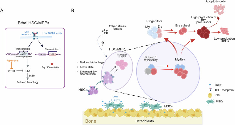
The hematopoietic stem cell and multipotent progenitor (HSC/MPP) pool dynamically responds to stress to adapt blood output to specific physiological demands. In β-thalassemia (Bthal), severe anemia and ineffective erythropoiesis generate expansion of erythroid precursors and a chronic stress status in the bone marrow (BM) microenvironment. However, the response to the BM altered status at the level of the HSC/MPP compartment in terms of lineage commitment has not been investigated. Bulk and single-cell RNA-sequencing reveal that Bthal HSCs/MPPs are expanded and activated with enhanced priming along the whole Ery differentiation trajectory. Consistently, HSC/MPP showed an altered TGFβ expression and autophagy transcriptional signatures along with a declined dormancy state. We discovered that the altered TGFβ signaling fosters the Ery potential of HSCs by reducing their autophagic levels, and in vivo stimulation of autophagy is sufficient to rescue the imbalance of the HSC compartment. Our findings identify the interplay between TGFβ and HSC autophagy as a key driver in the context of non-malignant hematopoiesis.


Conflict of interest statement

Competing interests: The authors declare no competing interests.

Figures

Fig. 1
Fig. 1. HSCs and MPPs characterization of bone marrow-derived CD34+ cells from thalassemic patients.
A Frequencies of LT-HSCs within BM CD34+ cells from Bthal patients, shown as fold relative to HD (HD n = 11; Bthal n = 9). mean ± SEM is shown. B Frequencies of MPPs within BM CD34+ cells from Bthal patients, shown as fold relative to HD (HD n = 11; Bthal n = 9). p-values by two-tailed unpaired t-test is shown; mean ± SEM is shown. C Frequencies of Ery-enriched subsets in the progenitor compartment (CMP and MEP), identified using CD71 and BAH1 as markers, within BM CD34+ cells from Bthal patients compared to HDs (HD n = 4; Bthal n = 10). p-values by two-tailed unpaired t-test is shown. mean ± SEM is shown. D Principal Component Analysis (PCA) performed on the 500 most significant Differential Expression Genes (DEGs) in Bthal versus HD hematopoietic subpopulations, by DESeq2 (HD n = 2; Bthal n = 2). E Heatmap of DEGs between Bthal and HD HSC/MPPs, by DESeq2, FDR < 0.001. F Volcano plot representation of DEGs in Bthal versus HD HSC/MPPs, by DESeq2, FDR < 0.05. G Gene Set Enrichment Analysis (GSEA) of categories from Hallmark MSigDB database in HSC/MPP pool of Bthal samples compared to HD, FDR < 0.05. Gene list of Subset1 (Ery/My/Ly) reported in Belluschi, S. et al. was also evaluated. FDR < 0.05. NES, normalized enrichment score. H GSEA of categories from REACTOME database in HSC/MPP pool of Bthal samples compared to HD, FDR < 0.05. I Differentially expression of selected genes (normalized UMI counts) involved in HSC maintenance between Bthal (red bars) and HD HSCs (white bars), (HD n = 2; Bthal n = 2). Results are represented as Fold respect to Ctrl; mean ± SEM is shown. Source data are provided as Source Data file.
Fig. 2
Fig. 2. Erythroid signature in primitive hematopoietic cells in Bthal.
Normalized UMI counts of selected erythroid-megakaryocytes genes differentially expressed between Bthal (red) and HD (grey), in HSCs (A) and MPPs (B) (HD n = 2; Bthal n = 2). Results are represented as Fold respect to Ctrl. mean ± SEM is shown. C Frequencies of Subset1 and Subset2 within HSC/MPP pool in HD and Bthal samples (HD n = 3; Bthal n = 3). mean ± SEM is shown. D Frequencies of Subset1 and Subset2 within HSC CD49f+ cells in HD and Bthal samples (HD n = 3; Bthal n = 3). mean ± SEM is shown. E Cloning efficiency of the HSC/MPP Subset 1 of Bthal samples compared to HD ones. Mean/number of colonies/2000 cells/plate (each dot represents the mean of 2 technical replicates) from Bthal and HD HSC/MPP Subset plated in a CFU assay (n = 8 from HD n = 3; n = 10 from Bthal n = 3). mean ± SEM is shown. F Percentage of colonies generated by Bthal and HD HSC/MPP Subset1 and containing differentiated cells (HD n = 3; Bthal n = 3). The type of colony: erythroid (E), granulocyte and myeloid (GM) or a combination of both (mix) is shown. P-value by two-tailed unpaired t-test; mean ± SEM is shown. G Cloning efficiency of the HSC/MPP Subset 2 of Bthal samples compared to HD ones. Mean/number of colonies/2000 cells/plate (each dot represents the mean of 2 technical replicates) from Bthal and HD HSC/MPP Subset plated in a CFU assay (n = 5 from HD n = 3; n = 8 from Bthal n = 3). p-value by two-tailed unpaired t-test; mean ± SEM is shown. H Percentage of colonies generated by Bthal and HD HSC/MPP Subset2 and containing differentiated cells (HD n = 3; Bthal n = 3). The type of colony: erythroid (E), granulocyte and myeloid (GM) or a combination of both (mix) is shown. mean ± SEM is shown. Source data are provided as Source Data file.
Fig. 3
Fig. 3. Single cell landscape of CD34+ differentiation in Bthal.
Analysis of 10x Genomics scRNA-seq from CD34+ HSPCs and phenotypic HSC/MPP isolated from BM of Bthal patients or HD. A UMAP for 36,878 single cells from 6 Bthal patients and 6 HDs. Indicated lineage groups were derived from the conflation of clusters with similar cell identities annotated based on label transfer from Zeng et al. (HSC, MPP, MEP, Meg precursors, MEP, Erythroid, EoBasoMast precursor, Cycling prog, GMP, Mono, MDP, MLP, CLP, B cell precursor). Original cluster annotation is shown in Fig. S7A. B Bar graph of the relative composition of HSPC groups shown in A in Bthal and HD. P values by two-tailed unpaired t-test; mean ± SEM is shown. C Top panels: UMAPs colored by the expression of a dormant HSC signature (left) and CDK6 gene expression (right). Bottom panels: Gaussian kernel density estimates plotted in the UMAP for HD (left) and Bthal (right) CD34+ cells on UMAP. D Ridge plot of pseudotime values assigned to phenotypic HSC/MPPs of Bthal and HD. P-value by two-side Welsh t-test. Selected pathways E MSigDB hallmark, F published genesets related to stemness and lineage commitment) significantly enriched (padj<0.05) by GSEA in the populations indicated on the y axis. Red: enriched in Bthal; Blue: enriched. A list of all enriched pathways is provided in Supplementary Data 6, 7. Terms with adjusted p.value (Benjamini-Hochber correction) less than 0.05 were considered significantly enriched. Representative examples of genes differentially expressed along the Ery (G) or monocytic (H) differentiation trajectory as identified by tradeSeq. Purple lines: Bthal; yellow lines: HD. Source data are provided as Source Data file.
Fig. 4
Fig. 4. TGFβ inhibition induces erythroid priming of HSCs.
Normalized UMI counts of selected genes, involved in TGFβ (A) and BMP (B) signaling, differentially expressed between Bthal HSCs (red) and HD HSCs (white). Results are represented as Fold respect to Ctrl (HD n = 2; Bthal n = 2). mean ± SEM is shown. C Transcript level of molecules involved in TGFβ and BMP signaling evaluated by ddPCR in MSCs. P value by two-tailed unpaired t-test; mean ± SEM is shown. D Evaluation of TGFβ1 in Bthal and HD platelet-poor plasma by enzyme-linked immunosorbent assay (ELISA) (Bthal n = 11; HD n = 9). P value by two-tailed unpaired t-test; mean ± SEM is shown. E Experimental plan for in vivo treatment of humanized mice with TGFβi inhibitor and/or BMP2. CD34+ mPB cells were administered to NBSGW female mice by retro-orbital injection. 9- to 10-week-old NBSGW mice were conditioned with busulfan injected intraperitoneally (15 mg/kg body weight) 24 h before transplantation. After 10 weeks, mice were treated with 10 mg/kg of TGFβ inhibitor (TGFβi) by subcutaneous injection and/or 20 ng/g of BMP2 molecule by intraperitoneal injection, twice weekly for 2 weeks. Image was created with BioRender.com. (https://BioRender.com/3tbmj37). F Frequencies of hCD45+ in BM from untreated animals (ctrl n = 8) and after in vivo treatment with TGFβi inhibitor (n = 8 mice), with BMP2 (n = 6 mice), or the combination of both (TGFβi + BMP2 n = 10 mice), from n = 2 independent experiments. P-values by two-tailed unpaired t-test; mean ± SEM is shown. G Frequencies of CD34+ in hCD45+ BM from untreated animals (ctrl n = 8) and after in vivo treatment with TGFβi inhibitor (n = 8 mice), with BMP2 (n = 6 mice), or the combination of both (TGFβi + BMP2 n = 10 mice), from n = 2 independent experiments. P-values by two-tailed unpaired t-test; mean ± SEM is shown. H Frequencies of HSC CD49f+ in hCD34+ from untreated animals (ctrl n = 8) and after in vivo treatment with TGFβi inhibitor (n = 8 mice), with BMP2 (n = 6 mice), or the combination of both (TGFβi + BMP2 n = 10 mice), from n = 2 independent experiments. P-values by two-tailed unpaired t-test; mean ± SEM is shown. I Frequencies of Subset2 in untreated animals (ctrl n = 8) and after in vivo treatment with TGFβi inhibitor (n = 8 mice), with BMP2 (n = 6 mice), or the combination of both (TGFβi + BMP2 n = 10 mice), from n = 2 independent experiments. P-values by two-tailed unpaired t-test; mean ± SEM is shown. J Frequencies of Subset 1 in untreated animals (ctrl n = 8) and after in vivo treatment with TGFβi inhibitor (n = 8 mice), with BMP2 (n = 6 mice), or the combination of both (TGFβi + BMP2 n = 10 mice), from n = 2 independent experiments. P-values by two-tailed unpaired t-test; mean ± SEM is shown. K Subset1/Subset2 ratio in all groups of treated mice relative to untreated ones. Group of mice: TGFβi inhibitor (n = 8 mice), with BMP2 (n = 6 mice), or the combination of both (TGFβi + BMP2 n = 10 mice), from n = 2 independent experiments. P-values by two-tailed unpaired t-test; mean ± SEM is shown. L Colony assay on CD34+ cells purified from the untreated and treated animals. The percentage of BFUe colonies is shown. Each dot represents the mean of two dishes/mouse. Untreated animals (ctrl n = 4), TGFβi inhibitor (n = 8), BMP2 (n = 6), or the combination of both (TGFβi + BMP2 n = 5), from n = 2 independent experiments. P-values by two-tailed unpaired t-test; mean ± SEM is shown. Source data are provided as a Source Data file.
Fig. 5
Fig. 5. Autophagy is reduced in Bthal LT-HSCs.
A DEGs carrying BMP responsive element (BMP-RE) between Bthal and HD HSC/MPP. Gene Ontology (GO) analysis of 120 target genes of BMP pathways, padj <0.01. Terms with adjusted p-value (Benjamini-Hochber correction). B GSEA of categories from MSigDB database in HSC/MPP pool of Bthal samples compared to HD, FDR < 0.05. C Genes of autophagy deregulated in Bthal HSC. Normalized UMI counts of selected genes, involved in Autophagy, differentially expressed between Bthal HSCs (red) and HD HSCs (white). Results are represented as Fold respect to Ctrl (HD n = 2; Bthal n = 2). D Representative microscopy images of DAPI (blue) and LC3 staining (green) of purified HD and Bthal sorted LT-HSCs. Scale bar 10 μm. n = 3 independent experiments (n = 3 HD samples and n = 3 Bthal samples). E Quantification of LC3 median intensity of evaluated foci in HD and Bthal LT-HSCs (HD n = 3; Bthal n = 3). About total 100 nuclei were analyzed for condition. P-values by two-tailed unpaired t-test; mean ± SEM. F the area of evaluated foci in HD and Bthal LT-HSCs (46 nuclei from HD n = 3; 32 nuclei from Bthal n = 3). P-values by two-tailed unpaired t-test; mean ± SEM. Source data are provided as Source Data file.
Fig. 6
Fig. 6. TGFβ inhibition induces a reduction of autophagic activity.
A Normalized UMI counts of selected genes, differentially expressed between th3/+ HSCs (red) and wt HSCs (blue) purified by FACS. Results are represented as Fold respect to wt (th3/+ n = 3; wt n = 4). *p < 0.05, by two-tailed unpaired t-test; mean ± SEM is shown. B Normalized UMI counts of selected genes, involved in TGFβ signaling, differentially expressed between th3/+ HSCs (red) and wt HSCs (blue). Results are represented as Fold respect to wt (th3/+ n = 3; wt n = 4). *p < 0.05, **p < 0.01, by two-tailed unpaired t-test; mean ± SEM is shown. C Normalized UMI counts of selected genes, involved in autophagy pathway, differentially expressed between th3/+ HSCs (red) and wt HSCs (blue). Results are represented as Fold respect to wt. (th3/+ n = 3; wt n = 4). *p < 0.05, **p < 0.01, by two-tailed unpaired t-test; mean ± SEM is shown. D Percentage of Cyto-ID+ in LT-HSCs in wt (n = 10) and treated th3/+ mice (n = 11) from n = 2 independent experiments. p-value by two-tailed unpaired t-test; mean ± SEM is shown. E Evaluation of TGFβ1 in th3/+ and wt BM fluid by ELISA (wt n = 8; th3/+ n = 8). p-value by two-tailed unpaired t-test; mean ± SEM is shown. F Experimental plan for in vivo treatment of wt mice with TGFβi inhibitor. Image created with BioRender.com. (https://BioRender.com/ho2ypan). G Frequencies of LT-HSCs in untreated animals (ctrl n = 9) and after in vivo treatment with TGFβi inhibitor (n = 12), from n = 2 independent experiments; p-value by one-tailed unpaired t-test; mean ± SEM is shown. H Frequencies of MPPMk/E (in untreated animals (ctrl n = 9) and after in vivo treatment with TGFβi inhibitor (n = 12), from n = 2 independent experiments; p-value by two-tailed unpaired t-test; mean ± SEM is shown. I Klf1 expression measured on sorted LT-HSCs from ctrl (n = 5) and treated wt mice (n = 9) from n = 3 independent experiment. p-value by Mann-Whitney test; mean ± SEM is shown. J Percentage of Cyto-ID+ in LT-HSCs in ctrl wt (n = 5) and treated wt mice (n = 5). p-value by Mann-Whitney test. mean ± SEM is shown. K Map1lc3b gene expression measured on sorted LT-HSCs from ctrl (n = 9) and treated wt mice (n = 10). p < 0.0001 by two-tailed unpaired t-test; mean ± SEM is shown. The percentage of Cyto-ID+ in LT-HSCs (L) in untreated (n = 6) and after in vitro treatment of th3/+ LT-HSCs with TGFβ1 molecule (n = 6 samples) or Bafilomycin A (n = 6 samples), as control. Each sample is a pool of 4-6 mice. (n = 3 independent experiments). p-values by one-way ANOVA, with Dunnett’s multiple comparison test. Mean ± SEM is shown. M Quantification of Cyto-ID level dye levels (MFI) in untreated (n = 6 samples) and after in vitro treatment of th3/+ LT-HSCs with TGFβ1 molecule (n = 6 samples) or Bafilomycin A (n = 5 samples), as control. Values are shown as fold versus the untreated one. p-value by Mann-Whitney test. mean ± SEM is shown. N Frequencies of LT-HSCs, MPPMk/E in HSPC at 12–20 weeks after transplantation from th3/+ (n = 5) and Ulk1-/- th3/+ (n = 6) mice from n = 2 independent experiments P-values by two-tailed unpaired t-test; mean ± SEM is shown. O Gene expression of Klf1 was measured on sorted LT-HSCs from th3/+ (n = 5) and Ulk1-/- th3/+ (n = 6) mice from n = 2 independent experiments. Relative expression on HPRT. th3/+, red; Ulk1-/- th3/+ green. Two-tailed unpaired t-test; mean ± SEM is shown. P Gene expression of Klf1 was measured on sorted MPPMk/E (P from th3/+ (n = 5) and Ulk1-/- th3/+ (n = 6) mice from n = 2 independent experiments. Relative expression on HPRT. th3/+, red; Ulk1-/- th3/+ green. Two-tailed unpaired t-test; mean ± SEM is shown. Source data are provided as Source Data file.
Fig. 7
Fig. 7. mTOR inhibition restores the unbalanced hematopoiesis in Bthal.
A Experimental plan for in vivo treatment of wt and th3/+ mice with rapamycin 4 mg/kg i.p. Image created with Biorender. (https://BioRender.com/qli1427). B Frequencies of HSC in untreated animals and after in vivo treatment with mTOR inhibitor rapamycin. wt (n = 9) mice, blue; th3/+ (n = 12) mice, red; th3/+ Rapa (n = 17) mice, wt Rapa (n = 9) mice from 3 independent experiments. p-values by Mann-Whitney test; mean ± SEM is shown. C Frequencies of MPPMK/E in untreated animals and after in vivo treatment with mTOR inhibitor rapamycin. wt (n = 9) mice, blue; th3/+ (n = 12) mice, red; th3/+ Rapa (n = 17) mice, wt Rapa (n = 9) mice from 3 independent experiments. p-values by Mann-Whitney test; mean ± SEM is shown. D Gene expression of KLF1 measured on sorted LT-HSCs from wt (n = 9) mice, blue; th3/+ (n = 7) mice, red; th3/+ Rapa (n = 9) mice, orange; wt Rapa (n = 5) mice, from 3 independent experiments. p-values by Mann-Whitney test; mean ± SEM is shown. E Gene expression of KLF1 measured on sorted MPP MK/E from wt (n = 5) mice, blue; th3/+ (n = 10) mice, red; th3/+ Rapa (n = 10) mice, orange; wt Rapa (n = 4) mice from 3 independent experiments. Relative expression on HPRT. P-values by Mann-Whitney test; mean ± SEM is shown. F Percentage of Cyto-ID+ in LT-HSCs in untreated th3/+ (n = 9) and th3/+ Rapa LT-HSCs (n = 7). P-values by two-tailed unpaired t-test; mean ± SEM is shown. G Quantification of CYTO-ID dye levels (MFI) in untreated th3/+ (n = 9) and th3/+ Rapa LT-HSCs (n = 7). p-value by Mann-Whitney test; mean ± SEM is shown. H pS6 levels measured by flow cytometry in untreated (n = 6) and treated th3/+ LT-HSCs (n = 7). p < 0.0001 by two-tailed unpaired t-test; mean ± SEM is shown. I Cloning efficiency of the HSC/MPP Subset1of HD Ctrl and treated with Rapamycin (5 ng/ml). Number of colonies/2000 cells/plate from HD HSC/MPP Subset1 plated in a CFU assay with or without rapamycin (n = 3 independent experiments; each dot represents the mean of 2 technical replicates). mean ± SEM is shown. J Percentage of colonies generated by HSC/MPP Subset1 Ctrl and in response to treatment with Rapamycin (n = 3 independent experiments). The type of colony: erythroid (E), granulocyte and myeloid (GM) or a combination of both (mix) is shown. P-value two-way ANOVA, Šídák’s multiple comparisons test, mean ± SEM is shown. Source data are provided as Source Data file.
Fig. 8
Fig. 8. Model of hematopoiesis in β-thalassemia.
In β-thalassemia, rare multipotent (Ery/My/Ly) primitive cells are leaving their quiescent state and shifting towards an active status with a preferential erythroid lineage differentiation. A) Specifically, inhibition of TGFβ signaling leads LT-HSC towards erythroid lineage by increasing Klf1 expression and by reducing the autophagy, in terms of Map1lc3b gene expression and mTOR activation. Rapamycin treatment restores the hematopoietic process by inducing autophagic activity in LT-HSCs. B). Low TGFβ1 production in Bthal BM niche by MSCs and OBs causes a reduction of autophagy in HSC, favoring the loss of stemness and an acceleration towards the erythroid lineage. A subset of cells with Ery potential can quickly differentiate in response to niche signals, thus depleting Ery cells from the progenitor fraction, or can be directly committed towards erythroid precursors. Other stress signals could contribute to the altered HSC cell fate in Bthal. TGFβ1-dependent autophagy in Bthal HSC/MPP cells. Based on the inhibitory strategy, TGFβ signaling regulates autophagy and erythroid commitment of primitive cells. The image was created with BioRender.com. (https://BioRender.com/99gtlee).

Similar articles

References

    1. Velten, L. et al. Human haematopoietic stem cell lineage commitment is a continuous process. Nat. Cell Biol.19, 271–281 (2017). - PMC - PubMed
    1. Akashi, K., Traver, D., Miyamoto, T. & Weissman, I. L. A clonogenic common myeloid progenitor that gives rise to all myeloid lineages. Nature404, 193–197 (2000). - PubMed
    1. Doulatov, S., Notta, F., Laurenti, E. & Dick, J. E. Hematopoiesis: a human perspective. Cell Stem Cell10, 120–136 (2012). - PubMed
    1. Busch, K. et al. Fundamental properties of unperturbed haematopoiesis from stem cells in vivo. Nature518, 542–546 (2015). - PubMed
    1. Haas, S., Trumpp, A. & Milsom, M. D. Causes and consequences of hematopoietic stem cell heterogeneity. Cell Stem Cell22, 627–638 (2018). - PubMed

Substances

LinkOut - more resources