. 2025 Jul 1;16(1):5460.
doi: 10.1038/s41467-025-60289-0.

Autoregulation of the real-time kinetics of the human mitochondrial replicative helicase

Affiliations

Autoregulation of the real-time kinetics of the human mitochondrial replicative helicase

Ismael Plaza-G A et al. Nat Commun. .

Abstract

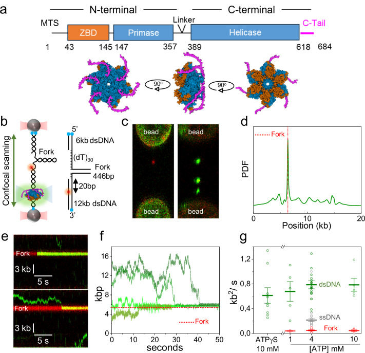
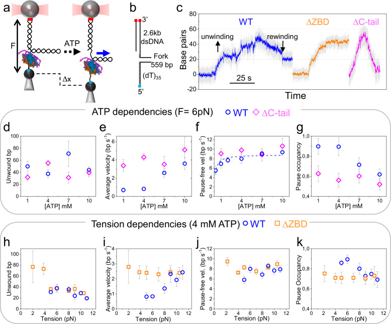
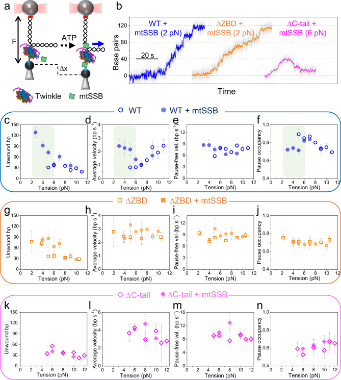
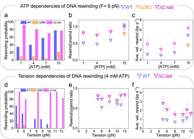
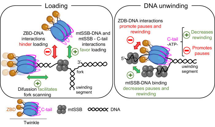
The human mitochondrial helicase Twinkle is essential for mitochondrial DNA (mtDNA) replication and integrity. Using biochemical and single-molecule techniques, we investigated Twinkle's real-time kinetics, including DNA loading, unwinding, and rewinding, and their regulation by its N-terminal Zinc-binding domain (ZBD), C-terminal tail, and mitochondrial SSB protein (mtSSB). Our results indicate that Twinkle rapidly scans dsDNA to locate the fork, where specific interactions halt diffusion. During unwinding, ZBD-DNA interactions and C-terminal tail control of ATPase activity downregulate kinetics, slowing down the helicase. Binding of mtSSB to DNA likely outcompetes ZBD-DNA interactions, alleviating the downregulatory effects of this domain. Furthermore, we show that ZBD-DNA interactions and ATP binding also regulate rewinding kinetics following helicase stalling. Our findings reveal that ZBD and C-terminal tail play a major role in regulation of Twinkle´s real-time kinetics. Their interplay constitutes an auto-regulatory mechanism that may be relevant for coordinating the mtDNA maintenance activities of the helicase.


Conflict of interest statement

Competing interests: The authors declare no competing interests.

Figures

Fig. 1
Fig. 1. Loading and diffusion of Twinkle on DNA.
a (Top) Twinkle consists of an ancestral N-terminal primase domain and a C-terminal helicase domain, connected by a linker region. Residues (numbers) corresponding to the Zinc Binding Domain (ZBD, orange) and the carboxyl-terminal tail (C-Tail, magenta) were deleted in the ΔZBD and ΔC-tail variants, respectively. MTS-Mitochondrial Targeting Sequence. (Bottom) Representation of Twinkle hexamer, with ZBD in orange and the C-tail in magenta, prepared by superimposing AlfaFold2 predicted monomers with T7gp4 hexamer (PDB 1E0K). b (Left) Diagram of the optical tweezers-confocal assay to image eGFP-Twinkle (green) on individual DNA constructs containing a DNA fork attached between optically trapped beads (Methods). (Right) Schematics of the fork DNA construct showing the helicase loading site, (dT)30, the distance of the fluorophore to the junction (20 bp) and the lengths of the unwinding segment (446 bp) and dsDNA handles labeled with biotin (blue dots). c Example scans showing separately (left) the Atto 647N-labelled fork position (red) and (right) Twinkle diffraction-limited spots (green) on the DNA construct. d Distribution of initial positions of eGFP-Twinkle diffraction-limited spots on the DNA constructs showing preferential binding at the fork position (N = 50, PDF considers the error in the position of each oligomer as the standard deviation). e Representative kymographs of eGFP-Twinkle oligomers (green) binding directly (top) or diffusing before loading (bottom) at the fork position (in red). f Representative position vs. time plots of eGFP-Twinkle spots show that diffusion is halted upon finding the fork position (4 mM ATP). g Diffusion coefficients of individual Twinkle units (6 ± 2 monomers) on dsDNA (green, N = 39 independent Twinkle units), ssDNA (grey, N = 38, Supplementary Fig. 4) and DNA fork (red, N = 21) at different ATP concentrations or in the presence of ATPγS. Error bars represent standard error of the mean (s.e.). For this figure (f, g) source data are provided as a Source Data file.
Fig. 2
Fig. 2. ATP and tension dependencies of real-time DNA unwinding kinetics.
a Schematic of optical trapping assays. A DNA fork-like construct is tethered between two functionalized micron-sized beads. One bead is hold in the optical trap (red cone), while the other is immobilized on a micropipette. In the presence of ATP, DNA unwinding is recorded as a change in tether extension (Δx) under constant mechanical tension (F). The blue arrow indicates the 5′ to 3′ translocation of the helicase along the hairpin. b The DNA construct consists of a 559 bp DNA hairpin (sequence in Supplementary Methods) with a ∼2.6-kb long dsDNA handle labelled with digoxigenin (red dots) and a 5′-poly(dT)35 labeled with biotin (blue dot). The single stranded poly(dT)35 tail serves as a helicase loading site (Methods). c Representative traces of the WT Twinkle (blue), ΔZBD (orange) and ΔC-tail (magenta) variants (F = 6 pN). DNA unwinding (increase in bp) is often followed by rewinding (decrease in bp). Raw data (grey background) was smoothed using a low-pass 10 Hz filter. dg ATP dependencies of the average unwinding processivity (d), unwinding rate (e), pause-free velocity (f), and pause occupancy (g) for the WT helicase (blue), and ΔC-tail (magenta) variant. The dotted line in (f) represents the Michaelis-Menten fit for the pause-free velocity of WT Twinkle as a function of ATP concentration (R2 = 0.89). Data in (d, g) were recorded at F = 6 pN, the minimal tension enabling consistent activity detection across all ATP concentrations (WT, N = 46 independent activities; Δ-Ctail, N = 36). hk Tension dependencies of the average unwinding processivity (h), unwinding rate (i), pause-free velocity (j) and pause occupancy (k) for WT Twinkle (blue, N = 45) and the ΔZBD variant (orange, N = 32). For all panels, data points represent to the average of multiple independent measurements, and error bars indicate the s.e. For this figure (dk) source data are provided as a Source Data file.
Fig. 3
Fig. 3. Tension dependencies of real-time DNA unwinding kinetics in the presence of mtSSB.
a Schematic of optical trapping assays with mtSSB. The experimental setup is identical to that described in Fig. 2a, with the addition of mtSSB (5 nM, green shape) in the reaction buffer. Experiments were performed at 4 mM ATP. b Representative unwinding traces with mtSSB. Traces show the activity of WT Twinkle (blue, F = 2 pN), ΔZBD (orange, F = 2 pN) and ΔC-tail (magenta, F = 6 pN) variants in the presence of mtSSB. Under these conditions, only the ΔC-tail exhibited significant rewinding events upon unwinding. Raw data (grey background) was smoothed using a low-pass 10 Hz filter. c–f Tension dependencies of the average unwinding processivity (c), unwinding rate (d), pause-free velocity (e) and pause occupancy (f) for WT Twinkle in the absence (blue empty symbols, N = 45) and presence (solid blue symbols, N = 57) of mtSSB. The shaded box highlights the tension range where mtSSB’s stimulatory effects are most apparent. g–j panels show same metrics as above (cf) for the ΔZBD variant in the absence (empty orange symbols, N = 32) and presence (full orange symbols, N = 29) of mtSSB. k–n panels show same metrics as above (cf) for the ΔC-tail variant in the absence (empty magenta symbols, N = 27) and presence (solid magenta symbols, N = 18) of mtSSB. For all panels, data points represent the average of multiple independent measurements, and error bars indicate the s.e. For this figure (cn) source data are provided as a Source Data file.
Fig. 4
Fig. 4. ATP and tension dependencies of DNA rewinding events.
a Bar plots shows the probability of rewinding events following unwinding for the WT helicase (blue bars, N = 46), ΔZBD (orange bars, N = 25), and ΔC-tail (magenta bars, N = 36) at varying ATP concentrations. b Average rewinding/unwinding ratios for the WT (blue symbols, N = 17), ΔZBD (orange symbol, N = 4) and ΔC-tail (magenta symbols, N = 29) as a function of ATP concentration. Ratios ~1 indicate that the number of rewound bp is similar to the number unwound bp. Data for the ΔZBD variant is shown only at 10 mM ATP due to insufficient rewinding events at lower ATP concentrations for meaningful analysis. c Average rewinding velocities for the WT (blue symbols, N = 20), ΔZBD (orange symbol, N = 4) and ΔC-tail (magenta symbols, N = 25) variants as a function of ATP concentration. d Probabilities of rewinding events following unwinding for the WT helicase (blue bars, N = 45), ΔZBD (orange bars, N = 32) and ΔC-tail (magenta bars, N = 27) variants at varying mechanical tension. e Average rewinding/unwinding ratios for the WT (blue symbols, N = 15), and ΔC-tail (magenta symbols, N = 24) variant as a function of tension. f Average rewinding velocities for the WT (blue symbols, N = 16) and ΔC-tail (magenta symbols, N = 23) variant under varying tension. ATP dependencies were measured at 6 pN and tension dependencies were measured at 4 mM ATP. Data points represent the average of multiple independent measurements, and error bars indicate the s.e. For this figure (bf) source data are provided as a Source Data file.
Fig. 5
Fig. 5. Autoregulation of Twinkle activities by the ZBD and C-terminal tail.
Twinkle scans DNA to locate the fork junction through diffusion. In the absence of mtSSB, ZBD-DNA interactions restrict functional loading at the fork and promote pauses (and rewinding events) during DNA unwinding. During this reaction, the C-tail downregulates ATP turnover, leading to additional pauses and reduced DNA unwinding rate. In the presence of mtSSB, interactions between the mtSSB and the C-terminal tail facilitate functional loading of the helicase onto the fork. mtSSB binding to the translocation strand outcompetes the inhibitory ZBD-DNA interactions, enhancing functional loading, decreasing pause occupancy during DNA unwinding, and reducing the probability of rewinding events.

Similar articles

References

    1. Spelbrink, J. N. et al. Human mitochondrial DNA deletions associated with mutations in the gene encoding Twinkle, a phage T7 gene 4-like protein localized in mitochondria. Nat. Genet.28, 223–231 (2001). - PubMed
    1. Peter, B. & Falkenberg, M. TWINKLE and other human mitochondrial DNA helicases: structure, function and disease. Genes11, 408 (2020). - PMC - PubMed
    1. Sen, D., Patel, G. & Patel, S. S. Homologous DNA strand exchange activity of the human mitochondrial DNA helicase TWINKLE. Nucleic Acids Res.44, 4200–4210 (2016). - PMC - PubMed
    1. Sen, D., Nandakumar, D., Tang, G.-Q. & Patel, S. S. Human mitochondrial DNA helicase TWINKLE is both an unwinding and annealing helicase. J. Biol. Chem.287, 14545–14556 (2012). - PMC - PubMed
    1. Khan, I. et al. Biochemical characterization of the human mitochondrial replicative twinkle helicase: SUBSTRATE SPECIFICITY, DNA BRANCH MIGRATION, AND ABILITY TO OVERCOME BLOCKADES TO DNA UNWINDING. J. Biol. Chem.291, 14324–14339 (2016). - PMC - PubMed

MeSH terms

LinkOut - more resources