. 2025 Jul 1;16(1):5688.
doi: 10.1038/s41467-025-61206-1.

Generating combinatorial diversity via engineered V(D)J-like recombination in Saccharomyces cerevisiae

Affiliations

Generating combinatorial diversity via engineered V(D)J-like recombination in Saccharomyces cerevisiae

Andrew P Cazier et al. Nat Commun. .

Abstract

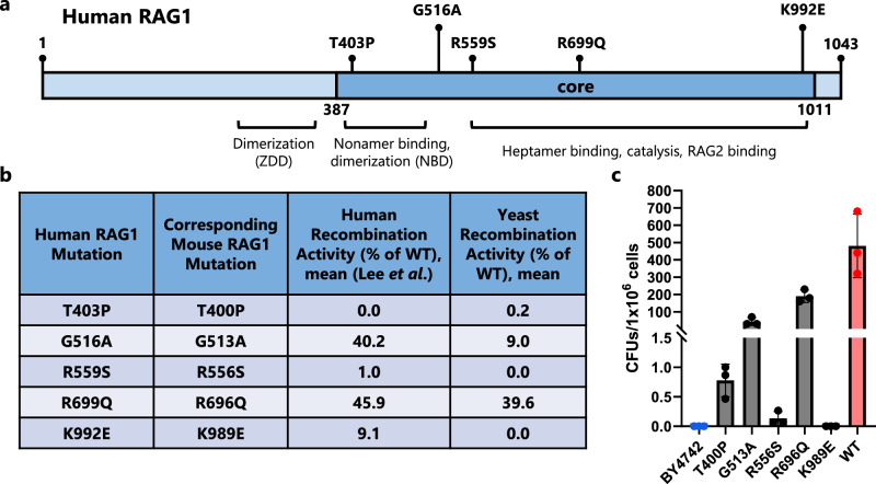
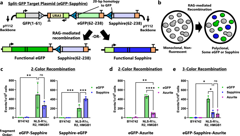
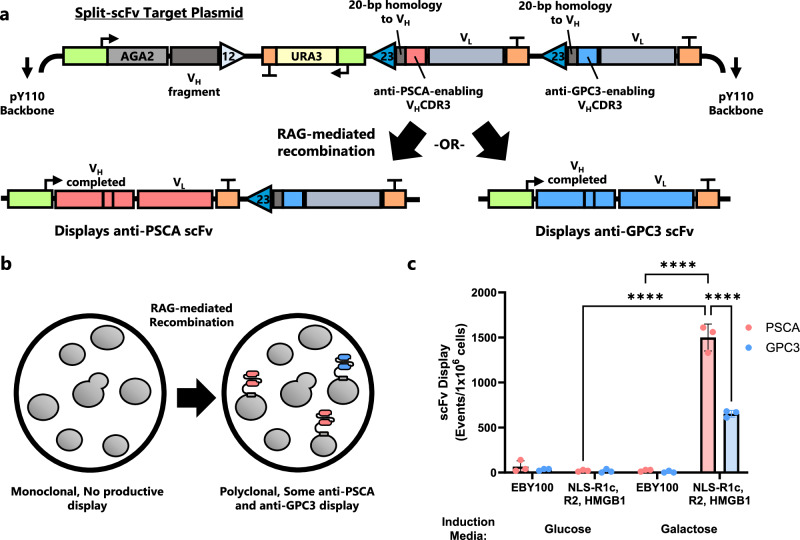
V(D)J recombination is integral to the development of antibody diversity and proceeds through a complex DNA cleavage and repair process mediated by several proteins, including recombination-activating genes 1 and 2, RAG1 and RAG2. V(D)J recombination occurs in all jawed vertebrates but is absent from evolutionarily distant relatives, including the yeast Saccharomyces cerevisiae. As yeast grow quickly and are a platform for antibody display, engineering yeast to undergo V(D)J recombination could expand their applicability for studying antibody development. Therefore, in this work we incorporate RAG1 and RAG2 into yeast and characterize the resulting recombination ability using a split antibiotic resistance assay, demonstrating successful homology-assisted formation of coding joints. By pursuing a variety of strategies, we increase the rate of homology-assisted recombination by over 7000-fold, with the best rates approaching 1% recombination after four days. We further show that our platform can assay the severity of several disease-causing RAG1 mutations. Finally, we use our engineered yeast to simultaneously generate up to three unique fluorescent proteins or two distinct antibody fragments starting from an array of nonfunctional gene fragments, which we believe to be the first-ever generation of genetic and phenotypic diversity solely using random recombination of preexisting DNA in a non-vertebrate cell.


Conflict of interest statement

Competing interests: The Authors declare the following competing interests: J.B. and A.P.C. have filed a patent related to this work. The other authors declare no competing interests.

Figures

Fig. 1
Fig. 1. eGFP tagging of RAG1 and RAG2 sheds light on expression and nuclear localization.
a RAG1 or RAG2 variants were tagged with eGFP on the C-terminus and integrated in BY4742-NAB2-mCherry yeast. Median eGFP fluorescence was then measured using flow cytometry after overnight culture in YPG. b Confocal microscopy images of eGFP-tagged RAG1 and RAG2 protein variants (green) in a NAB2-mCherry strain which natively highlights the nucleus (red). Scale bar represents 5 μm; all images are at the same scale. Gains have been adjusted from the original images to aid visual comparison. The brightfield channel for each image can be found in Supplementary Fig. S1. c Nuclear localization was estimated by calculating the Pearson correlation coefficient between the RAG-eGFP and NAB2-mCherry signals of the confocal images. d Confocal microscopy images of eGFP-tagged RAG1 protein variants (green) in a NOP56-mCherry strain which natively highlights the nucleolus (red). As in (b), scale bar represents 5 μm. The complete set of images can be found in Supplementary Fig. S2. e Nucleolar localization was estimated by calculating the Pearson correlation coefficient between the RAG-eGFP and NOP56-mCherry signals of the confocal images. RFU = relative fluorescence units, NLS = nuclear localization signal. In (a, c, and e) data are presented as mean values ± SD; n = 3 biological replicates. Statistical significance was calculated with a one-way ANOVA and Tukey test (*p < 0.05, **p < 0.01, ***p < 0.001, and ****p < 0.0001). From left to right, the highlighted p values are, in (a), p = 0.0014, <0.0001, 0.0012, and 0.0001; in (c), p = <0.0001, 0.0499, 0.0016, and 0.0194; and in (e), p = <0.0001 and <0.0001. Source data are provided in the Source Data file.
Fig. 2
Fig. 2. Protein engineering increases activity of RAG-mediated, homology-assisted recombination of split antibiotic resistance gene.
a Diagram of pY112-CJA-UP-H20 recombination target plasmid. Triangles represent RSSs. Cutting by RAG1/2 leads to homology-assisted DNA repair of the G418R gene that confers resistance to the antibiotic G418. b Yeast strains were engineered with both RAG1 and RAG2 genes (either full-length, core, or core NLS variants) and optionally HMGB1. After 4-d induction in SG-Leu media, cell cultures were plated on Leu, G418 plates and colonies were counted after outgrowth. c G418R recombination with yeast strains assessing the combination of full-length RAG2 with RAG1core. Cells were plated after a 4-d induction. d G418R recombination with alternate truncations of RAG1 that include more regions of the full-length protein than the initial RAG1core construct (which contains amino acids 383-1006). All strains include full-length RAG2 and HMGB1. Cells were plated after a 4-d induction. C = core protein, F = full-length protein, N-C = core protein with N-terminal nuclear localization signal, NLS = nuclear localization signal, CFU = colony forming unit. In (bd) strains that appear in multiple plots are highlighted with a color, and data are presented as mean values ± SD; n = 3 biological replicates. Statistical significance was calculated with a one-way ANOVA and Tukey test (ns = not significant, **p < 0.01, and ****p < 0.0001). From left to right, the highlighted p values are, in (b), p = <0.0001 and 0.0042; in (c), p = <0.0001 and <0.0001; and in (d), p = <0.0001 and 0.0148. Source data are provided in the Source Data file.
Fig. 3
Fig. 3. Homology and intersignal sequence strongly influence G418R recombination assay.
a G418R recombination assay using substrates with mutated RSSs that cannot be recognized by the RAG proteins (Mut12 and Mut23) or nonstandard pairings (12 + 12 and 23 + 23). Cells were cultured for 4 d in SG-Leu prior to plating. b G418R recombination assay showing the effect of adjusting homology length between the two halves of the G418R gene in the recombination target plasmid. Cells were plated after an 8-d induction. c Diagram of the original intersignal region for “UP” plasmid design and the shorter variant sequences between the RSSs that were tested. For all alternatives, 20 bp of homology between the two halves of G418R was used. d G418R recombination assay varying the intersignal region between RSSs. See (c) for region descriptions. Cells were plated after a 4-d induction. e Similar to (d), G418R recombination assay testing additional intersignal regions between RSSs. Cells were plated after a 4-d induction. f G418R recombination assay using enhanced pY112-CJA-U-H20 plasmid with additional recombination strains. Recombination strains contained RAG2, HMGB1, and the specified variant of RAG1. Cells were plated after 4-d induction. In all figures, BY4742 are wild-type cells that do not contain V(D)J recombination genes. CFU = colony forming unit. In (a, b, df) data are presented as mean values ± SD; n = 3 biological replicates. In (a, d, and e), statistical significance was calculated using a two-way ANOVA and Tukey test. In (b), significance was calculated using multiple two-sided t-tests with a Holm-Sidak correction for multiple comparisons. In (f), statistical significance was calculated using a one-way ANOVA and Tukey test (ns = not significant, *p < 0.05, **p < 0.01, ***p < 0.001, ****p < 0.0001). From left to right, the highlighted p values are, in a, p = 0.9811, 0.9916, 0.9916, 0.7759, 0.9602, and <0.0001; in (b), p = 0.2606, 0.0078, 0.0028, and 0.0004; in (d) p = 0.0329, < 0.0001; in (e) p = 0.0017 and <0.0001; and in (f) p = 0.0001 and 0.1147. Source data are provided in the Source Data file.
Fig. 4
Fig. 4. Signal joint formation and isolation using an integrated target.
a Diagram of the integrated SJUE target in chromosome XV (Chr XV). Cutting by RAG1/2 can lead to NHEJ repair which fuses the RSSs together and forms a signal joint. Such an event permanently removes eGFP and URA3, leading to cells that do not fluoresce and are 5-FOA-resistant. b The SJUE target was integrated in both a recombination-competent (AC518: NLS-RAG1core, RAG2, and HMGB1) and recombination-incompetent (BY4742) strain. After an 8-d induction in YPG media, cells were plated on SD, 5-FOA plates. Both eGFP-positive and eGFP-negative colonies were separately counted. CFU = colony forming unit. In (b), data are presented as mean values ± SD; n = 3 biological replicates. Statistical significance was calculated using a two-way ANOVA with repeated measures across fluorescence types and Fisher LSD test. Source data are provided in the Source Data file.
Fig. 5
Fig. 5. Recombination efficiency of pathogenic RAG1 mutants in yeast correlates with activity in mammalian cells.
a Genetic map of human RAG1 showing the core region and the location of five mutations which have been shown to cause varying degrees of immunodeficiency. Certain domains of RAG1 and their function are highlighted, including the zinc dimerization domain (ZDD) and nonamer-binding domain (NBD). b Table of human RAG1 mutants and their corresponding mutation in mouse RAG1. Additionally, the previously reported activity of the mutants in mammalian cells is given along with the activity measured in yeast in this work. WT = wild-type RAG1. c G418R recombination with pY112-CJA-U-H20 plasmid. BY4742 are the base strain of yeast. All other strains contain mouse RAG1core with a mutation (or without in the case of wild type, WT), RAG2, and HMGB1. Cells were plated after a 4-d induction. CFU = colony forming unit. In (c), data are presented as mean values ± SD; n = 3 biological replicates. Source data are provided in the Source Data file.
Fig. 6
Fig. 6. RAG-mediated recombination of split-GFP construct generates diversity—up to three colors in vivo.
a Diagram of the pY112-CJCG-ES-H20 plasmid. The first portion of GFP is flanked by a 12-RSS followed by an intersignal region and then two modules each consisting of a 23-RSS, a gene fragment that creates eGFP or Sapphire, and a terminator. Triangles represent RSSs. Each gene fragment has 20 bp of homology to the first portion of GFP. Due to the 12/23 rule of RAG recombination, there are only two expected resolutions to recombination, one which creates eGFP and another which creates Sapphire. b Expected phenotype change for cells bearing the pY112-CJCG-ES-H20 plasmid as recombination is induced. Initially, all cells are non-fluorescent, but RAG-mediated recombination leads to subpopulations with eGFP or Sapphire fluorescence. c 2-color recombination of cells with target plasmids that have a fragment order of eGFP then Sapphire (pY112-CJCG-ES-H20) or Sapphire then eGFP (pY112-CJCG-SE-H20). d 2-color recombination with fragment order eGFP then Azurite (pY112-CJCG-EA-H20). e 3-color recombination using a target plasmid with a fragment order of eGFP, Sapphire, then Azurite (pY112-CJCG-ESA-H20). For all tests, cells were cultured for 9-d in SG-Leu media prior to analysis with flow cytometry. BY4742 are wild-type cells that do not contain V(D)J recombination genes. In (ce) data are presented as mean values ± SD; n = 3 biological replicates. Statistical significance was calculated with a two-way ANOVA with repeated measures across colors and Fisher LSD test (for c and d) or Tukey test (for e) (ns = not significant, *p < 0.05, **p < 0.01, ***p < 0.001, ****p < 0.0001). From left to right, the highlighted p values are, in (c), p = 0.0027, 0.1741, 0.0001, and 0.0001; in (d), p = 0.0012 and < 0.0001; and in (e), p = 0.0327, 0.8738, and 0.0136. Source data are provided in the Source Data file.
Fig. 7
Fig. 7. Two functional scFv variants can be generated with recombination and displayed.
a Diagram of the pY110-CJCS-PG-H20 plasmid. AGA2 and a VH fragment are flanked by a 12-RSS followed by an intersignal region and then two modules each consisting of a 23-RSS, a gene fragment that contains a unique VHCDR3, and a terminator. Triangles represent RSSs. RAG-mediated recombination creates either an anti-PSCA or anti-GPC3 product which can be displayed on the surface of the cell. b Demonstration of expected phenotype change during RAG-mediated recombination of the split-scFv substrate. Initially, the cells cannot display a functional scFv. Depending on the outcome, a fraction of cells will display either an anti-PSCA or anti-GPC3 antibody after recombination. c scFv recombination results measured via flow cytometry. Wild-type EBY100 cells were compared to the recombination strain which expresses NLS-RAG1core, RAG2, and HMGB1. Cells were induced for 8 d in SG-Trp (Galactose) or SD-Trp (Glucose), then grown for 1 d in SD-Trp followed by 1 d in buffered SG-Trp. Due to fluorophore overlap, the cells were split into two separate staining reactions: one for PSCA and Myc and another for GPC3 and FLAG. In (c) data are presented as mean values ± SD; n = 3 biological replicates. Statistical significance was calculated with a two-way ANOVA with repeated measures to compare scFv display and Tukey test (****p < 0.0001). Source data are provided in the Source Data file.

Similar articles

References

    1. Schatz, D. G. et al. The Mechanism, Regulation and Evolution of V(D)J Recombination. in Molecular Biology of B Cells (Third Edition) Chapter 2, (eds. Honjo, T., Reth, M., Radbruch, A., Alt, F. & Martin, A.) 13–57 (Academic Press, London, 2024).
    1. Rees, A. R. Understanding the human antibody repertoire. mAbs12, 1729683 (2020). - PMC - PubMed
    1. Qi, Q. et al. Diversity and clonal selection in the human T-cell repertoire. Proc. Natl Acad. Sci.111, 13139–13144 (2014). - PMC - PubMed
    1. Briney, B., Inderbitzin, A., Joyce, C. & Burton, D. R. Commonality despite exceptional diversity in the baseline human antibody repertoire. Nature566, 393–397 (2019). - PMC - PubMed
    1. DeKosky, B. J. et al. Large-scale sequence and structural comparisons of human naive and antigen-experienced antibody repertoires. PNAS113, E2636–E2645 (2016). - PMC - PubMed

MeSH terms

LinkOut - more resources