. 2025 Jul 1;15(1):21204.
doi: 10.1038/s41598-025-04286-9.

Optogenetic restoration of high-sensitivity vision using ChRmine- and ChroME-based channelrhodopsins

Affiliations

Optogenetic restoration of high-sensitivity vision using ChRmine- and ChroME-based channelrhodopsins

Victoria C Fong et al. Sci Rep. .

Abstract

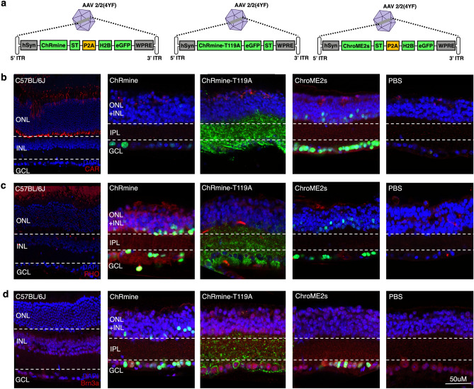
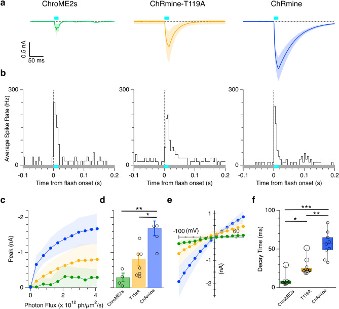
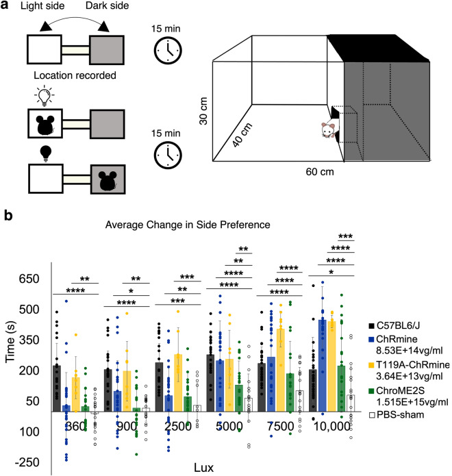
Optogenetic gene therapy is a promising mutation-independent treatment that aims to restore visual perception in patients blinded by retinal diseases that cause photoreceptor degeneration. Still, low sensitivity or slow kinetics of currently utilized optogenetic proteins limit the efficacy of such approaches. Here, we evaluated the therapeutic potential of three channelrhodopsin variants: ChRmine, from the algae Rhodomonas lens, ChRmine-T119A, a faster-closing ChRmine variant, and ChroME2s, a second-generation Chronos-based opsin.We expressed these opsins in retinal ganglion cells of rd1 mice, a model of severe retinal degeneration. Single cell electrophysiology demonstrates opsin's large sensitivity to a range of light intensities as well as opsin-expressing retinal ganglion cells generated action potentials in response to light stimulation. Behavioral tests showed ChRmine-T119A's efficacy at 360 lux compared to unmodified ChRmine and ChroME2s. ChRmine and ChroME2s did restore light perception at higher light intensities. Additionally, our dose-response study with ChRmine-T119A revealed that lower viral titers were more effective at restoring light sensitivity. Our study demonstrates that these ChRmine- and ChroME-based opsins can enhance vision in late-stage blinding diseases.

Keywords: Channelrhodopsin; Electrophysiology; Gene therapy; Mouse behavior; Optogenetics; Retinal disease; Vision restoration.


Conflict of interest statement

Declarations. Competing interests: The authors declare no competing interests. Ethics statement: The animal study was approved by the Animal Care and Use Committee (ACUC) at University of California, Berkeley. The study was conducted in accordance with the local legislation and institutional requirements.

Figures

Fig. 1
Fig. 1
Immunohistochemistry of retinas treated with ChRmine, ChRmine-T119A, and ChroME2s AAVs. (a) Schematic of the AAV transgene cassettes. (b-d) Immunofluorescence stained-images of retinal sections. eGFP represents opsin expression in treated rd1 mice. Cone photoreceptors are labeled using CAR (b, red) and rod photoreceptors with RHO (c, red). (d) Co-localization of the opsin (eGFP) and Brn3a (red) confirm expression in ganglion cells. CAR = cone arrestin, RHO = rhodopsin, eGFP = enhanced green fluorescent protein.
Fig. 2
Fig. 2
Characterization of RGCs expressing ChroME2s, ChRmine-T119A, and ChRmine opsins in response to blue LED light flashes. (A) Average ChroME2s (n = 3), ChRmine (n = 4), and ChRmine-T119A (n = 4 cells) mediated photocurrents in RGCs in response to diffuse 485 nm light flash of 167 mW/cm2 (timing shown by the cyan bar). (B) Average peristimulus spike-time histograms (PSTH) generated from ChroME2s- (200.0 ± 102.5 Hz, n = 12 cells), ChRmine-T119A- (152.4 ± 144.2 Hz, n = 14 cells), and ChRmine- (152.9 ± 84.2 Hz during stimulus, n = 17 cells) expressing RGCs. Light flash was 32.2 mW/cm2 for 15 ms. (C) Average peak photocurrent as a function of flash intensity. (D) Max peak photocurrent at the highest light intensity for ChRmine- (1.69 ± 0.42 nA, n = 4 cells), ChRmine-T119A- (0.81 ± 0.47 nA, n = 7 cells), and ChroME2s-expressing RGCs (0.30 ± 0.24 nA, n = 4 cells). Error bars in bar graph are represented as SEM (E) Current–voltage relations for the peak photocurrents of ChroME2s- (n = 36 cells), ChRmine-T119A- (n = 7 cells), and ChRmine-expressing RGCs (n = 6 cells). (E) Exponential decay time constants measured from the photocurrents in ChroME2s- (9.5 ± 8.2, n = 8 cells), ChRmine-T119A- (25.6 ± 9.3 ms, n = 12 cells), and ChRmine-expressing RGCs (56.2 ± 16.5 ms, n = 9 cells). *p < 0.05, **p < 0.01, and ***p < 0.001. Shading shows the standard deviation in all panels.
Fig. 3
Fig. 3
Light avoidance experiments in rd1 mice expressing ChRmine, ChRmine-T119A, or ChroME2s. (a) Schematic of light/dark box for light avoidance test based on Riebe and Wotjak, 2012. (b) Average change in side preference using white light (irradiance of 100 μW cm−2) in C57BL6/J (n = 26), ChRmine- (n = 29), ChRmine-T119A- (n = 8), ChroME2s- (n = 21), and PBS- (n = 19) injected mice. Statistical significance assessed with Welch’s ANOVA test with post-hoc Games-Howell HSD threshold matrix, *p < 0.05, **p < 0.01, ***p < 0.0001.
Fig. 4
Fig. 4
Light aversion experiments in rd1 mice treated with different titers of ChRmine-T119A. Mice treated with lower titers are more light-sensitive than mice treated with the highest titer. Average change in side preference using white light (irradiance of 100 μW cm−2). C57BL/6J (n=26), ChRmine-T119A undiluted (n = 18), ChRmine-T119A 1:2 (n = 8), ChRmine-T119A 1:5 (n = 7), ChRmine-T119A 1:10 (n = 7), and PBS (n = 19). Data represents the mean ± SD. Statistical significance assessed with Welch’s ANOVA test with post-hoc Games-Howell HSD threshold matrix, *p < 0.05, **p < 0.01, ***p < 0.0001.

References

    1. Ku, C. A. & Pennesi, M. E. The new landscape of retinal gene therapy. Am. J. Med. Genet. C Semin. Med. Genet.184, 846–859 (2020). - PubMed
    1. Botto, C. et al. Early and late stage gene therapy interventions for inherited retinal degenerations. Prog. Retin. Eye Res.86, 100975 (2022). - PubMed
    1. Stefanov, A. & Flannery, J. G. A systematic review of optogenetic vision restoration: History, challenges, and new inventions from bench to bedside. Cold Spring. Harb. Perspect. Med.13, a041154 (2023). - PMC - PubMed
    1. Pfeiffer, R. L. & Jones, B. W. Current perspective on retinal remodeling: Implications for therapeutics. Front. Neuroanat.16, 1099348 (2022). - PMC - PubMed
    1. Puthussery, T. & Taylor, W. R. Functional changes in inner retinal neurons in animal models of photoreceptor degeneration. Adv. Exp. Med. Biol.664, 525–532 (2010). - PubMed

MeSH terms

Substances