. 2025 Jul 1;16(1):469.
doi: 10.1038/s41419-025-07793-9.

Neuroprotection of photoreceptors by combined inhibition of both Fas and autophagy pathways in P23H mice

Affiliations

Neuroprotection of photoreceptors by combined inhibition of both Fas and autophagy pathways in P23H mice

Mengling Yang et al. Cell Death Dis. .

Abstract

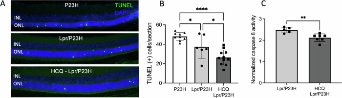
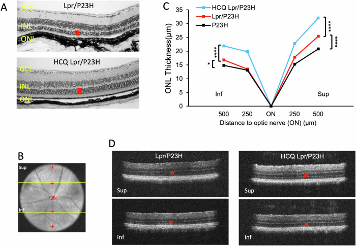
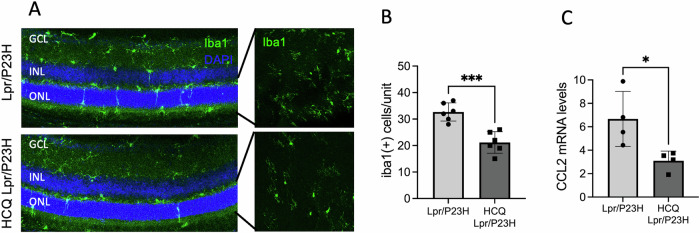
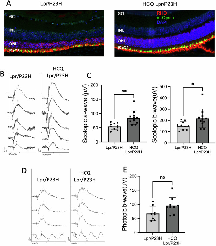
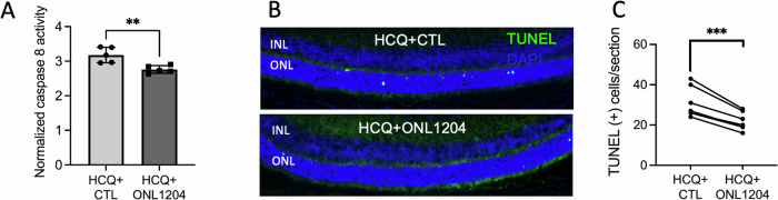
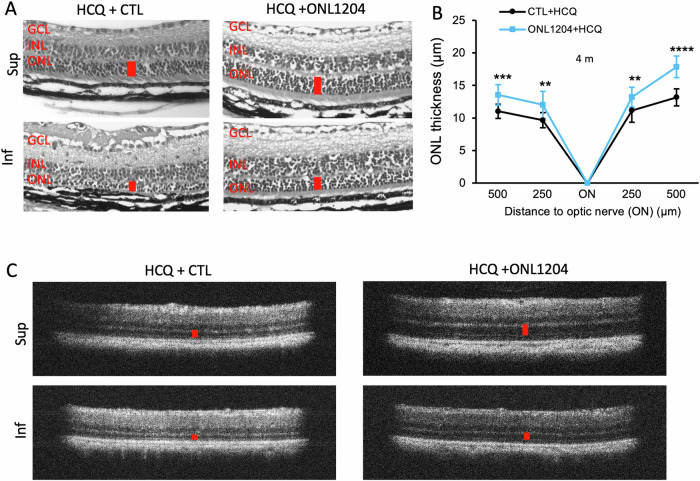
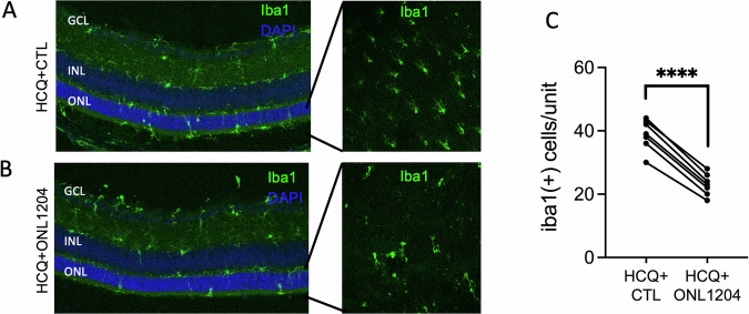
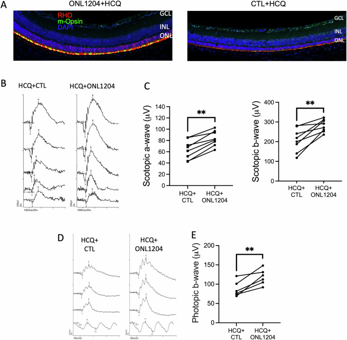
The P23H variant of rhodopsin (RHO) is a common cause of autosomal dominant retinitis pigmentosa (adRP). Our previous data have shown that both the Fas (CD95) death receptor and hyperactivation of autophagy contribute to photoreceptor (PR) death in a mouse model of P23H-RHO adRP. Individually, inhibition of Fas or suppression of autophagy flux improves PR survival and function. The purpose of this study is to examine whether combined inhibition of Fas receptor activation and reducing autophagy flux would have an additive effect on PR survival and function in the P23H mouse. We crossed the Lpr mouse (which contains a functional knockout of the Fas receptor) with the P23H mouse to generate the Lpr/P23H mouse. Hydroxychloroquine (HCQ) was given in the drinking water at P21 to reduce autophagy flux. As an alternative to genetic inhibition of the Fas receptor, pharmacological blockade of the Fas receptor was achieved using intravitreal injections of the Fas inhibitor, ONL1204, administered via intravitreal injection at P14 and 2 months of age. Fellow eyes were injected with vehicle solution as controls. PR cell death, structure and function of the retina, as well as the activation of immune cells, were evaluated. Consistent with previous data, the Lpr/P23H mice exhibited a decreased rate of photoreceptor degeneration and reduced inflammation compared with P23H. Treatment of these mice with HCQ further preserved photoreceptor survival and function lowered the activation of immune cells, and resulted in reduced production of inflammatory cytokines in the retina. These results were recapitulated in HCQ-treated P23H mice receiving intravitreal injections of ONL1204. Our data suggest that in the mouse model of P23H adRD, inhibition of both the Fas pathway and autophagy pathways results in a greater protective effect, demonstrating the potential multipronged therapeutic approach to reduce PR death and improve retinal function in patients with P23H.


Conflict of interest statement

Competing interests: Two of the authors have relevant disclosures. AJK is an employee of ONL Therapeutics, the company that produces ONL1204. DNZ is an employee of the University of Michigan and is also a co-founder of ONL Therapeutics. DNZ also holds patents through the University of Michigan that are licensed to ONL Therapeutics. DNZ laboratory receives grant funding from ONL Therapeutics. All other authors have no competing interests. Ethics approval: All animal experiments and experimental methods were performed in accordance with the guidelines set forth by the University of Michigan Animal Care Committee and conformed to the Use of Animals in Ophthalmology and Vision Research established by the Association for Research in Vision and Ophthalmology (ARVO).

Figures

Fig. 1
Fig. 1. Decreased photoreceptor cell death and inflammation in the HCQ-treated Lpr/P23H mice.
A Representative TUNEL staining images and B quantification of TUNEL-positive cells in the ONL for P23H, Lpr/P23H mice, and HCQ-Lpr/P23H mice at P30 (n = 9 for P23H; n = 6 for Lpr/P23H and n = 11 for HCQ-Lpr/P23H. One-way ANOVA.). C Quantification of caspase 8 activity at 1 month of age (n = 7 for HCQ-treated Lpr/P23H, n = 6 for untreated Lpr/P23H). *P < 0.05, **P < 0.01; ****P < 0.0001. t-test. INL inner nuclear layer, ONL outer nuclear layer.
Fig. 2
Fig. 2. HCQ treatment preserved ONL thickness.
A Representative H&E staining images show preserved outer nuclear layer thickness (red bar) in the retina of HCQ-treated Lpr/P23H and untreated Lpr/P23H control mice. B Representative fundus photo showing the measurement points (red cross) in the superior and inferior of the retina by OCT. C Quantification of the thickness of the ONL (indicated by red cross in B) of the superior and inferior retina measured at both 250 and 500 µm from the optic nerve head by OCT in HCQ treated Lpr/P23H, Lpr/P23H, and P23H mice at the age of 4 months (n = 16 for HCQ Lpr/P23H and Lpr/P23H and n = 12 for P23H). D Representative OCT images of superior (Sup) and inferior (Inf) retina (indicated by yellow lines in B) of 4-month-old HCQ treated Lpr/P23H and Lpr/P23H controls. *P < 0.05; ****P < 0.0001, 1-way ANOVA. GCL ganglion cell layer, INL inner nuclear layer, ONL.
Fig. 3
Fig. 3. Decreased inflammation in the HCQ-treated Lpr/P23H mice.
A Representative images of the inferior area of the retinal whole mount and 30 µm retinal sections of HCQ-treated Lpr/P23H and untreated Lpr/P23H mice stained with Iba1 at 1 month of age. B Quantification of Iba1-positive cells in the ONL and subretinal space of the inferior retina of HCQ-treated Lpr/P23H and untreated Lpr/P23H mice (n = 6) paired t-test. C mRNA levels of inflammatory cytokines CCL2 in the retinas of HCQ-treated Lpr/P23H, untreated Lpr/P23H normalized to age-matched C57 mice at 1 month of age (n = 4) t-test and one-way ANOVA. *P < 0.05, ***P < 0.001. GCL gangling, INL inner nuclear layer, ONL outer nuclear layer.
Fig. 4
Fig. 4. HCQ treatment improved retinal function in Lpr/P23H mice.
A Representative immunostaining images of the retina of HCQ Lpr/P23H, Lpr/P23H, and C57 mice at 4 months of age, stained with rhodopsin (RHO in red), m-opsin (green), and DAPI (blue). B Representative scotopic traces (at 0.01, 1, 10, 32, and 64 cd s/m2) and C quantification of amplitudes of scotopic a-wave and b-wave of HCQ treated Lpr/P23H (n = 11), untreated Lpr/P23H mice (n = 10) at 4 months of age. D Representative photopic ERG traces (at 10, 32, and 100 cd s/m2) and E quantification of amplitudes of photopic b-wave of HCQ treated Lpr/P23H and control Lpr/P23H mice at 4 months of age. ns, not significant, *P < 0.05; **P < 0.01; one-tailed Wilcoxon test. GCL ganglion cell layer, INL inner nuclear layer, ONL outer nuclear layer, IS inner segment, OS outer segment.
Fig. 5
Fig. 5. In P23H mice, combining HCQ treatment with intravitreal injections of ONL1204 reduced photoreceptor death than HCQ treatment alone.
A Quantification of caspase 8 activity in the retina of HCQ + ONL1204 treated and HCQ+ Vehicle-treated eyes of the P23H mice at 1 month of age, normalized to ONL thickness (n = 10, 2 retinas from 2 mice were pooled for each sample). B Representative TUNEL staining images and C quantification of TUNEL-positive cells in the ONL for HCQ + ONL1204 treated and HCQ+ Vehicle-treated P23H eyes at 1 month (n = 6). **P < 0.01; ***P < 0.001, t-test. INL inner nuclear layer, ONL outer nuclear layer.
Fig. 6
Fig. 6. Combination of HCQ with ONL1204 preserved photoreceptors in the retinas of P23H mice.
A Representative H&E staining images show preserved ONL thickness (red bar) in the retinas of HCQ + ONL1204-treated eyes at 4 months of age. B Quantification of the thickness of the ONL of the superior and inferior retina measured at both 250 and 500 µm from the optic nerve head by OCT in HCQ + ONL1204 treated eyes and HCQ + CTL vehicle-treated control eyes at the age of 4 months (n = 15). C Representative OCT images of superior (Sup) and inferior (Inf) retina of 4-month-old HCQ + ONL1204 treated eye and HCQ + CTL vehicle-treated fellow eyes. **P < 0.01; ***P < 0.001; ****P < 0.0001, unpaired t-test. GCL ganglion cell layer, INL inner nuclear layer, ONL outer nuclear layer.
Fig. 7
Fig. 7. Treatment of HCQ + ONL1204 reduced inflammation in the retinas of P23H mice.
A Representative immunostaining images of retinal sections and retinal whole mount from inferior retinas of HCQ + ONL1204 treated eyes and B HCQ + CTL treated fellow eyes at 1 month of age stained with Iba-1 and DAPI. C Quantification of Iba1-positive cells in the ONL and subretinal space of the inferior retina of HCQ + ONL1204 treated eyes and HCQ + CTL treated eyes at 1 month of age (n = 7). ****P < 0.0001. Paired t-test. GCL ganglion cell layer, INL inner nuclear layer, ONL outer nuclear layer.
Fig. 8
Fig. 8. HCQ + ONL1204 treatment preserved visual function in P23H mice.
A Representative immunostaining images of the inferior retina of HCQ + ONL1204 treated eyes and HCQ+ control vehicle-treated fellow eyes of the P23H mice, at 4 months of age, stained with rhodopsin (red), m-opsin (green), and DAPI (blue). B Representative scotopic traces (at 0.01, 1, 10, 32, and 64 cd s/m2) and C quantification of amplitudes of scotopic a-wave and b-wave of HCQ + ONL1204 treated and HCQ + CTL treated fellow eye at 4 months of age. D Representative photopic ERG traces (at 10, 32, and 100 cd s/m2) and E quantification of amplitudes of photopic b-wave of HCQ + ONL1204 and HCQ + CTL-treated fellow eyes of P23H mice at 4 months of age (n = 8), **P < 0.01; one-tailed Wilcoxon test. GCL ganglion cell layer, INL inner nuclear layer, ONL outer nuclear layer.

Similar articles

References

    1. RetNet. https://sph.uth.edu/retnet/.
    1. Ben-Yosef T. Inherited retinal diseases. Int J Mol Sci. 2022;23:13467 10.3390/ijms232113467. - PMC - PubMed
    1. Sohocki MM, Daiger SP, Bowne SJ, Rodriquez JA, Northrup H, Heckenlively JR, et al. Prevalence of mutations causing retinitis pigmentosa and other inherited retinopathies. Hum Mutat. 2001;17:42–51. - PMC - PubMed
    1. Daiger SP, Bowne SJ, Sullivan LS. Perspective on genes and mutations causing retinitis pigmentosa. Arch Ophthalmol (Chicago, IL: 1960). 2007;125:151–8. - PMC - PubMed
    1. Jensen R. Differential effects of antipsychotic drugs on contrast response functions of retinal ganglion cells in wild-type Sprague-Dawley rats and P23H retinitis pigmentosa rats. PLoS ONE. 2019;14:1–14. - PMC - PubMed

MeSH terms

LinkOut - more resources